Review
. 2020;11(1):102-117.
doi: 10.1080/19490976.2019.1602428. Epub 2019 Apr 29.

Serine-rich repeat proteins from gut microbes

Affiliations
Review

Serine-rich repeat proteins from gut microbes

Dimitrios Latousakis et al. Gut Microbes. 2020.

Abstract

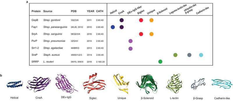
Serine-rich repeat proteins (SRRPs) have emerged as an important group of cell surface adhesins found in a growing number of Gram-positive bacteria. Studies focused on SRRPs from streptococci and staphylococci demonstrated that these proteins are O-glycosylated on serine or threonine residues and exported via an accessory secretion (aSec) system. In pathogens, these adhesins contribute to disease pathogenesis and represent therapeutic targets. Recently, the non-canonical aSec system has been identified in the genomes of gut microbes and characterization of their associated SRRPs is beginning to unfold, showing their role in mediating attachment and biofilm formation. Here we provide an update of the occurrence, structure, and function of SRRPs across bacteria, with emphasis on the molecular and biochemical properties of SRRPs from gut symbionts, particularly Lactobacilli. These emerging studies underscore the range of ligands recognized by these adhesins and the importance of SRRP glycosylation in the interaction of gut microbes with the host.

Keywords: Lactobacillus; Serine rich repeat protein; adhesin; gut symbiont; protein glycosylation.


Figures

Figure 1.
Figure 1.
Structural domain organization of characterized serine-rich repeat proteins (SRRPs) from gut commensal bacteria. (a) L. reuteri strain 100–23, (b) L. reuteri strain ATCC 53608, (c) Strep. salivarius strain JIM8777. SRRPs are composed of an N-terminal signal peptide (S; black boxes); an AST domain; a non-repeat domain that mediates adhesion (binding region; BR); two serine-rich repeat domains (SRR) flanking the BR (SRR-1 and SRR-2; checkered boxes); and a second non-repeat domain (N), followed by a C-terminal cell wall anchor domain (A; striped boxes). In contrast to the majority of SRRPs, SrpA from Strep. salivarius strains, including JIM8777, also harbors a number of MucBP (mucin-binding protein) domains before and/or after SRR-2 (nine after SRR-2 in the case of JIM8777; grey boxes). The SRRs are composed of serine residues alternating with, most frequently, either an alanine, valine, or threonine residue. Numbers represent the starting amino acid positions of each domain. White boxes represent non-repeat regions.
Figure 2.
Figure 2.
Organization of functionally characterized secA2/Y2 clusters from gut commensal bacteria. (a) L. reuteri 100–23, (b) L. reuteri ATCC 53608 and (c) Strep. salivarius JIM8777. The genes encoding the SecA2/Y2 translocation machinery are shown in red, the accessory secretion proteins Asp1-5 in blue and the priming GTs, GtfA, and GtfB in L. reuteri and GtfE and GtfF in Strep. salivarius, in yellow. Genes encoding additional GTs are shown in green and the genes encoding SRRPs are illustrated in teal. Black arrows represent genes that are not part of the aSec machinery. HP: hypothetical protein.
Figure 3.
Figure 3.
Circular phylogram representation of SRRP-BR domains from commensal and pathogenic bacteria. A MUSCLE multiple sequence alignment33 was carried out in MEGA-X34 using BR domain aa sequences from a total of 108 commensal-associated SRRPs and 32 pathogen/clinical-associated SRRPs. A guide tree was generated from the second iteration and a Maximum Likelihood phylogenetic tree displayed as a circular phylogram with EvolView software (http://www.evolgenius.info/evolview/).35 SRRP-BRs are displayed as follows: circle, from a commensal or non-pathogenic strain; star, from a pathogenic or clinical isolate. Host or source origins of strains are indicated as follows: pink, porcine; red, rodent; purple, human; aqua, avian; lime, bovine; blue, insect; yellow, sourdough; orange, other fermented food or drink; white, chimpanzee fruit residues. BRs shown with only a strain name are all from L. reuteri. The entries for which a SRRP-BR crystal structure is available are shaded in a light blue box. The scale bar represents the branch length expressed as the number of aa substitutions per site.
Figure 4.
Figure 4.
SRRP glycosylation mechanisms in gut commensal bacteria. (a) SRRP glycosylation in L. reuteri strains ATCC 53608 and 100–23. The GtfA/B complex initiates the glycosylation of the L. reuteri SRRP with GlcNAc residues, while GtfC extends the glycans with either GlcNAc (GtfC53608) or Glc (GtfC100–23). The glycosylated SRRP53608 is then secreted through the aSec system, whereas the SRRP100–23 is further extended by GtfD and/or GtfE before secretion. (b) SRRP glycosylation in Strep. salivarius strain JIM8777. SrpA, SrpB, and SrpC glycosylation are mediated by the GtfE/F complex, GTs encoded by a genetic locus unlinked to the aSec genomic island. After extension and acetylation of the glycans by other enzymes in the aSec system, the adhesins are secreted through the SecA2/Y2 channel. Blue circle: glucose, blue square: GlcNAc, SP: signal peptide.
Figure 5.
Figure 5.
Structural repertoire of the SRRP binding regions. (a) Summary of the domains found in the available structures of SRRPs. For each protein, we report the name, the source organism, the PDB code of the available structures, the year of the publication of the structure and the CATH code. On the right, a schematic representation is included of the structural domains present in the proteins. (b) Example of three-dimensional structure of each domain found in the SRRP-BRs.

References

    1. Lizcano A, Sanchez CJ, Orihuela CJ.. A role for glycosylated serine-rich repeat proteins in Gram-positive bacterial pathogenesis. Mol Oral Microbiol. 2012;27:257–269. doi:10.1111/j.2041-1014.2012.00653.x. - DOI - PMC - PubMed
    1. Seo HS, Xiong YQ, Sullam PM. Role of the serine-rich surface glycoprotein Srr1 of Streptococcus agalactiae in the pathogenesis of infective endocarditis. PLoS One. 2013;8:e64204. doi:10.1371/journal.pone.0064204. - DOI - PMC - PubMed
    1. Wu H, Mintz KP, Ladha M, Fives-Taylor PM. Isolation and characterization of Fap1, a fimbriae-associated adhesin of Streptococcus parasanguis FW213. Mol Microbiol. 1998;28:487–500. - PubMed
    1. Zhou M, Wu H. Glycosylation and biogenesis of a family of serine-rich bacterial adhesins. Microbiol (Reading, Engl). 2009;155:317–327. doi:10.1099/mic.0.025221-0. - DOI - PubMed
    1. Bensing BA, Sullam PM. An accessory sec locus of Streptococcus gordonii is required for export of the surface protein GspB and for normal levels of binding to human platelets. Mol Microbiol. 2002;44:1081–1094. - PubMed

Publication types

LinkOut - more resources