. 2019 Apr 29;16(1):13.
doi: 10.1186/s12977-019-0475-0.

Effect of transcription inhibition and generation of suppressive viral non-coding RNAs

Affiliations

Effect of transcription inhibition and generation of suppressive viral non-coding RNAs

Daniel O Pinto et al. Retrovirology. .

Abstract

Background: HIV-1 patients receiving combination antiretroviral therapy (cART) survive infection but require life-long adherence at high expense. In chronic cART-treated patients with undetectable viral titers, cell-associated viral RNA is still detectable, pointing to low-level viral transcriptional leakiness. To date, there are no FDA-approved drugs against HIV-1 transcription. We have previously shown that F07#13, a third generation Tat peptide mimetic with competitive activity against Cdk9/T1-Tat binding sites, inhibits HIV-1 transcription in vitro and in vivo.

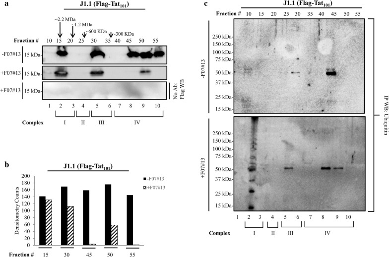
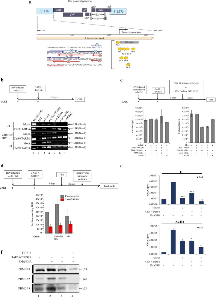
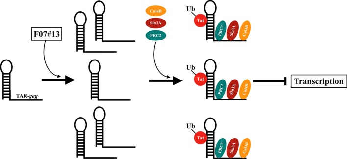
Results: Here, we demonstrate that increasing concentrations of F07#13 (0.01, 0.1, 1 µM) cause a decrease in Tat levels in a dose-dependent manner by inhibiting the Cdk9/T1-Tat complex formation and subsequent ubiquitin-mediated Tat sequestration and degradation. Our data indicate that complexes I and IV contain distinct patterns of ubiquitinated Tat and that transcriptional inhibition induced by F07#13 causes an overall reduction in Tat levels. This reduction may be triggered by F07#13 but ultimately is mediated by TAR-gag viral RNAs that bind suppressive transcription factors (similar to 7SK, NRON, HOTAIR, and Xist lncRNAs) to enhance transcriptional gene silencing and latency. These RNAs complex with PRC2, Sin3A, and Cul4B, resulting in epigenetic modifications. Finally, we observed an F07#13-mediated decrease of viral burden by targeting the R region of the long terminal repeat (HIV-1 promoter region, LTR), promoting both paused polymerases and increased efficiency of CRISPR/Cas9 editing in infected cells. This implies that gene editing may be best performed under a repressed transcriptional state.

Conclusions: Collectively, our results indicate that F07#13, which can terminate RNA Polymerase II at distinct sites, can generate scaffold RNAs, which may assemble into specific sets of "RNA Machines" that contribute to gene regulation. It remains to be seen whether these effects can also be seen in various clades that have varying promoter strength, mutant LTRs, and in patient samples.

Keywords: CRISPR; Gene silencing; HIV-1; Latency; Transcription; cART.


Conflict of interest statement

Authors declare no potential conflicts of interest. The content is solely the responsibility of the authors and does not necessarily represent the official views of the National Institutes of Health.

Figures

Fig. 1
Fig. 1
Effect of F07#13 on Tat degradation. a Following transfection into Jurkat cells, samples were collected and lysates were prepared for immunoprecipitation. Anti-Flag Ab was used for IP overnight, Protein A/G added the next day, washed, and samples were run on a gel and analyzed by Western blot for presence of Tat (α-Tat polyclonal Ab). Lanes 1 and 2 serve as control input transfected lysates (1/10) prior to IP. b Jurkat cells were transfected with 89.6 plasmid (20 μg) and CMV-Flag-Tat101 (20 μg) and 24 h later samples were treated with 0.01, 0.1, and 1 μM of F07#13 for an additional 48 h (a total 72 h). Cells were pelleted and washed, and lysates were run on a 4–20% Tris–glycine gel followed by Western blot with α-Flag antibody, followed by α-actin as a control. An IP with α-Flag antibody was run on a gel and probed with α-ubiquitin antibody. Densitometry was performed for each lane. c Cells were transfected with both 89.6 and Tat vector, followed by treatment with F07#13 (48 h; 1 μM) and two other inhibitors, MG132 (10 ng/mL) and a de-ubiquitin USP7 inhibitor (P5091; 3 μM), for 24 h and then separated on 4-20% Tris–glycine gel followed by Western blot with α-Flag antibody, α-ubiquitin antibody, and α-actin. Densitometry was performed to visualize changes in protein expression. The quantitation of 5 distinct bands in each lane was performed and summed to obtain total densitometry counts
Fig. 2
Fig. 2
Presence of ubiquitin-Tat in the large complex. a HIV-1 infected J1.1 cells were electroporated with CMV-Flag-Tat101 (20 μg) and kept at 37 °C for 48 h. Cells were isolated, washed, and extracts were processed for FPLC chromatography (Superose 6) using high salt. A total of 3.5 mg was used for chromatography. Flow rate parameters for the FPLC were set at 0.3 mL/min and 0.5 mL fractions of the flow-through were collected at 4 °C for approximately 60 fractions per sample (1 mL injected). Tat associated complexes were nanotrapped with NT084 and assayed for Western blot using α-Flag antibody. b Densitometry counts from panel a were obtained, normalized to background, and plotted to represent the relative abundance of Tat protein in each fraction. c Chromatography fractions were IPed with α-Flag antibody overnight, followed by addition of Protein A/G, ran on a gel, and analyzed by Western blot with α-ubiquitin antibody. Two sets of extracts (± F07#13) were run on chromatography and used for nanotrapping and Western blots
Fig. 3
Fig. 3
TAR-gag RNA association with various inhibitory complexes. a Early–mid log phase HIV-1 infected J1.1 cells were treated with F07#13 for 48 h (1 μM), pelleted, washed (×2) with PBS without Ca2+ and Mg2+, resuspended in lysis buffer, and 2500 µg of protein were equilibrated in degassed FPLC running buffer. A Superose 6 10/300 size-exclusion chromatography column was used to run lysed samples. Fractions were then pre-cleared with IgG for 2 h at 4 °C and then divided into 4 sub-fractions for IP using six antibodies against PSMD11, Sin3A, PRC2, HDAC-1, DNMT3A, and Cul4B (5 μg/reaction). Protein A/G was added the next day and the IPed complexes were washed. RNA was isolated for RT-qPCR using TAR-gag primers. An IP with IgG antibody was used as a control. Fractions from Complexes I, II, III, and IV constitute complex sizes from ~ 2.2 MDa to ~ 300 kDa. Error bars represent ± SD of three technical replicates. b Fractions from Complexes I, II, and III (500 µl) were nanotrapped with NT084 and assayed for RT-qPCR for presence of 7SK RNA. Fraction 10 was used as a control in lane 1 of this panel
Fig. 4
Fig. 4
Presence of HIV-1 RNA associated complexes in multiple HIV-1 infected whole cell extracts. a Fresh primary PBMCs (107 cells) were cultured with PHA/IL-2 for 7 days and infected with HIV-1 89.6 strain (MOI:1) [7]. Three days later they were treated with F07#13 (once every other day at 0.1 µM) for a total of 20 days. Cells were collected and lysates were loaded onto a sizing column under high salt. Column fractions were then IPed with antibodies against PRC2, Sin3A, Cul4B, and IgG. Following IP, RNA was isolated and samples were processed for RT-qPCR using primers against TAR-gag. Non-specific IgG background IPs were used as a control. Fractions from Complexes I, II, and III (500 µl) of infected PBMCs were nanotrapped with NT084 and assayed for RT-qPCR for presence of 7SK RNA (b) or half of the samples were run on an SDS/PAGE and Western blotted for presence of PRC2, Cul4B, actin, and Sin3A (data not shown) (c). Fraction 10 was used as a control in lane 1 of panels b and c. Error bars represent ± SD of three technical replicates
Fig. 5
Fig. 5
DNA-PK on the HIV-1 genome following Cas9+TAR3/6 transfection and alterations in cutting following F07#13 treatment. a Schematic of the HIV-1 proviral genome, which highlights the 5′ LTR of HIV-1. A series of gRNAs was designed to target the essential TAR loop required for Tat binding and proviral reactivation. b Three infected cell types (J1.1, CHME5/HIV, and U1) were grown in the presence of cART for 1 week prior to transfection. Cells were electroporated with three constructs at a 1:10 ratio (0.1 µg/1 µg of Cas9+TAR3/6) and kept in culture for 5 days. Approximately 1 × 107 cells were used for ChIP assay using antibodies (10 µg) against Pol II large subunit, Cdk9 (T186), p-H2AX, DNA-PK, and ARIDA. Following DNA purification, samples were PCR amplified using LTR primers and run on a 2% Agarose gel. c Similar to panel b except cells were treated with two inhibitors after 5 days. Both inhibitors, DNA-PK inhibitor (Nu 7441, 0.2 µM) and ATM inhibitor (KU 55933, 1 µM), were used for a 2 day treatment of either uninfected (Jurkat) or infected (J1.1) cells prior to CellTiter-Glo. Positive control Fas antibody was used for apoptosis on both cell types. d Similar experimental design to panel b, except J1.1, CHEM5/HIV, and U1 cells were treated with 100 nM TSA after 5 days of transfection. Viruses were isolated from the supernatants with NT086 particles and added to TZM-bl-Luc cells. e A similar experiment as outlined in panel d; however, U1 and ACH2 cells were treated 1 day prior to PHA/PMA treatment with either F07#13 (Day 4), Cas9+TAR3/6, or both together and analyzed by RT-qPCR for the presence of TAR RNA. *p value ≤ 0.05; ***p value ≤ 0.001. f Latent PBMCs (3 independent donors) were created as described previously [7]. After cART/IL-7 addition, samples were divided into 4 sections; two were electroporated (210 V) with TAR3/6 DNA ± F07#13 and kept in culture for 4 days. They were then treated with PMA/PHA for 2 days prior to p24 Western blot
Fig. 6
Fig. 6
A proposed model of the effect of F07#13 on binding to TAR-gag. The model is based on the notion that the ncRNAs (i.e. TAR-gag) are made from HIV-1 LTR and upon the introduction of F07#13, there is an increase in the copy number of TAR-gag due to non-processive Pol II transcription. The increased abundance of TAR-gag leads to the sequestration of ubiquitinated Tat, potentially through the TAR sequence. The presence of protein complexes with RNA can constitute newly synthesized “RNA machines”, which cause the repression of HIV-1 transcription through epigenetic modifications and potentially contribute to gene silencing and latency

References

    1. Akpamagbo YA, DeMarino C, Pleet ML, Schwab A, Rodriguez M, Barclay RA, Sampey G, Iordanskiy S, El-Hage N, Kashanchi F. HIV-1 transcription inhibitors increase the synthesis of viral non-coding RNA that contribute to latency. Curr Pharm Des. 2017;23:4133–4144. doi: 10.2174/1381612823666170622101319. - DOI - PubMed
    1. Ali A, Raja R, Farooqui SR, Ahmad S, Banerjea AC. USP7 deubiquitinase controls HIV-1 production by stabilizing Tat protein. Biochem J. 2017;474:1653–1668. doi: 10.1042/BCJ20160304. - DOI - PubMed
    1. Ammosova T, Berro R, Jerebtsova M, Jackson A, Charles S, Klase Z, Southerland W, Gordeuk VR, Kashanchi F, Nekhai S. Phosphorylation of HIV-1 Tat by CDK2 in HIV-1 transcription. Retrovirology. 2006;3:78. doi: 10.1186/1742-4690-3-78. - DOI - PMC - PubMed
    1. Angelov D, Charra M, Seve M, Côté J, Khochbin S, Dimitrov S. Differential remodeling of the HIV-1 nucleosome upon transcription activators and SWI/SNF complex binding. J Mol Biol. 2000;302:315–326. doi: 10.1006/jmbi.2000.4069. - DOI - PubMed
    1. Archin NM, Sung JM, Garrido C, Soriano-Sarabia N, Margolis DM. Eradicating HIV-1 infection: seeking to clear a persistent pathogen. Nat Rev Microbiol. 2014;12:750–764. doi: 10.1038/nrmicro3352. - DOI - PMC - PubMed

Publication types

Substances