. 2019 Apr 29;21(1):54.
doi: 10.1186/s13058-019-1136-x.

The opposing effects of interferon-beta and oncostatin-M as regulators of cancer stem cell plasticity in triple-negative breast cancer

Affiliations

The opposing effects of interferon-beta and oncostatin-M as regulators of cancer stem cell plasticity in triple-negative breast cancer

Mary R Doherty et al. Breast Cancer Res. .

Abstract

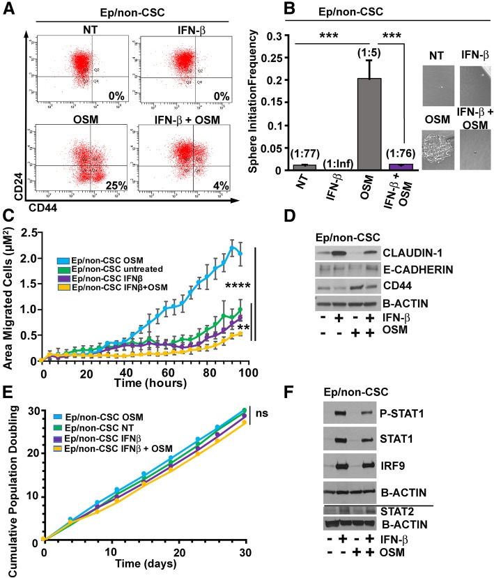
Background: Highly aggressive, metastatic and therapeutically resistant triple-negative breast cancers (TNBCs) are often enriched for cancer stem cells (CSC). Cytokines within the breast tumor microenvironment (TME) influence the CSC state by regulating tumor cell differentiation programs. Two prevalent breast TME cytokines are oncostatin-M (OSM) and interferon-β (IFN-β). OSM is a member of the IL-6 family of cytokines and can drive the de-differentiation of TNBC cells to a highly aggressive CSC state. Conversely, IFN-β induces the differentiation of TNBC, resulting in the repression of CSC properties. Here, we assess how these breast TME cytokines influence CSC plasticity and clinical outcome.

Methods: Using transformed human mammary epithelial cell (HMEC) and TNBC cell models, we assessed the CSC markers and properties following exposure to OSM and/or IFN-β. CSC markers included CD24, CD44, and SNAIL; CSC properties included tumor sphere formation, migratory capacity, and tumor initiation.

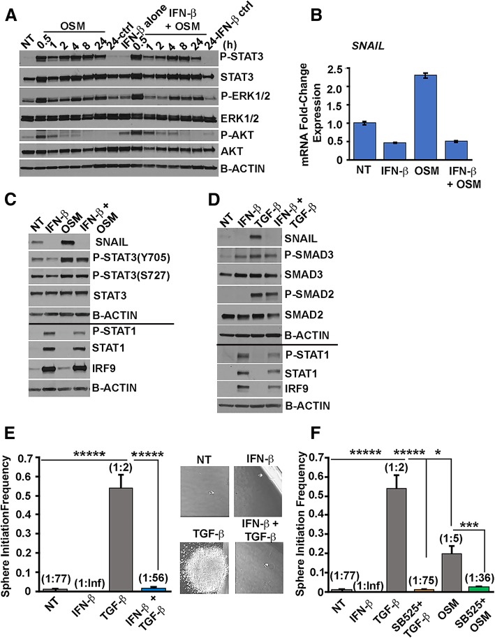
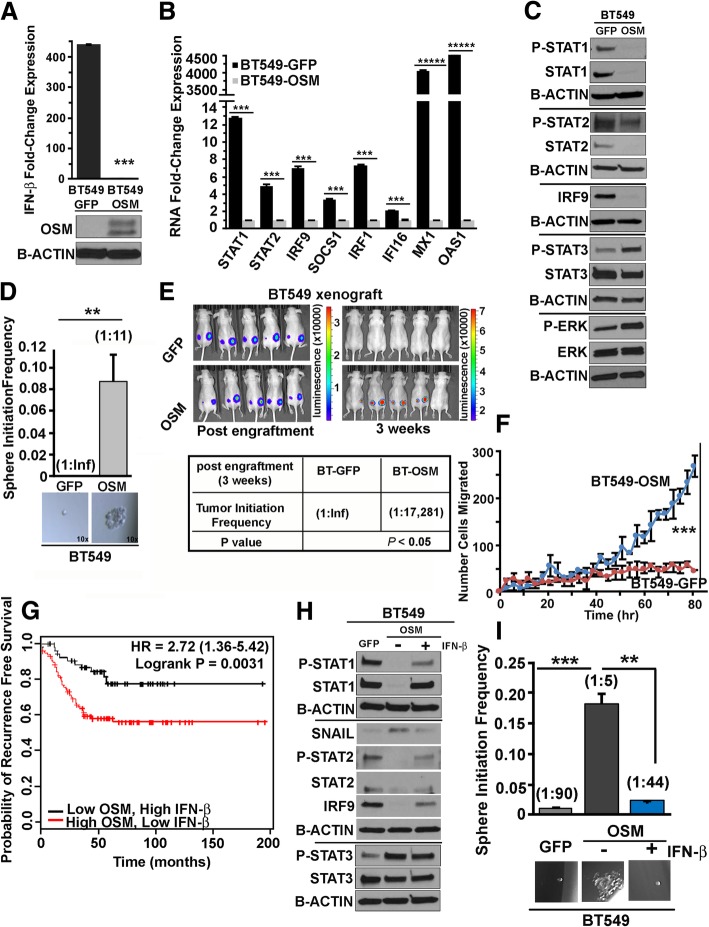
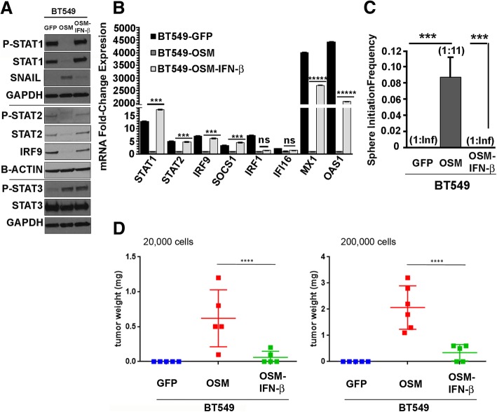
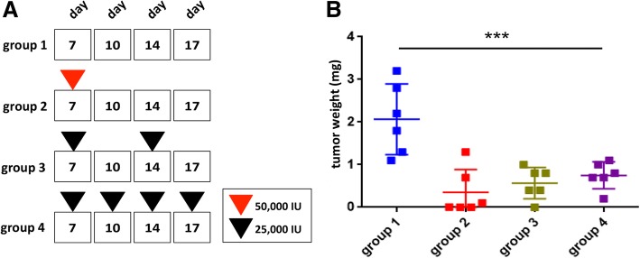
Results: There are three major findings from our study. First, exposure of purified, non-CSC to IFN-β prevents OSM-mediated CD44 and SNAIL expression and represses tumor sphere formation and migratory capacity. Second, during OSM-induced de-differentiation, OSM represses endogenous IFN-β mRNA expression and autocrine/paracrine IFN-β signaling. Restoring IFN-β signaling to OSM-driven CSC re-engages IFN-β-mediated differentiation by repressing OSM/STAT3/SMAD3-mediated SNAIL expression, tumor initiation, and growth. Finally, the therapeutic use of IFN-β to treat OSM-driven tumors significantly suppresses tumor growth.

Conclusions: Our findings suggest that the levels of IFN-β and OSM in TNBC dictate the abundance of cells with a CSC phenotype. Indeed, TNBCs with elevated IFN-β signaling have repressed CSC properties and a better clinical outcome. Conversely, TNBCs with elevated OSM signaling have a worse clinical outcome. Likewise, since OSM suppresses IFN-β expression and signaling, our studies suggest that strategies to limit OSM signaling or activate IFN-β signaling will disengage the de-differentiation programs responsible for the aggressiveness of TNBCs.

Keywords: Cancer stem cell plasticity; Cancer stem cells; Interferon-beta; Oncostatin-M; SNAIL; Triple-negative breast cancer; Tumor microenvironment.


Conflict of interest statement

Ethics approval and consent to participate

Experiments involving animals were performed in compliance with the guidelines approved by the Case Western Reserve University Institutional Animal Care and Use Committee (IACUC).

Consent for publication

Not applicable

Competing interests

The authors declare that they have no competing interests.

Publisher’s Note

Springer Nature remains neutral with regard to jurisdictional claims in published maps and institutional affiliations.

Figures

Fig. 1
Fig. 1
Sustained IFN-β exposure represses oncostatin-M-mediated cancer stem cell properties and inhibits migration. a Sustained exposure to IFN-β (Ep/non-CSC pre-treated with IFN-β (100 IU/mL) for 48 h prior to co-treatment with OSM (10 ng/mL) and IFN-β (100 IU/mL); co-treatments for 2 weeks) represses OSM-mediated CD44 acquisition, as shown by flow cytometry (top left, 0% CD44 in NT; top right, 0% CD44 in IFN-β alone; bottom left, 18% CD44 in OSM alone; bottom right, 4% CD44 in IFN-β + OSM co-treatment). b Sustained IFN-β exposure (Ep/non-CSC pre-treated with IFN-β (100 IU/mL) for 48 h prior to co-treatment with OSM (10 ng/mL) with IFN-β (100 IU/mL); co-treatments for 3 weeks) significantly represses OSM-mediated tumor sphere initiation at limiting dilution (stem cell frequency: 1:77 for control, 1:Inf (Infinity) for IFN-β, 1:5 for OSM alone, and 1:76 for IFN-β + OSM co-treatment, ***P < 0.0001) ± SD, n = 5. c Sustained IFN-β (Ep/non-CSC pre-treated with IFN-β (100 IU/mL) for 48 h prior to co-treatment with OSM (10 ng/mL) with IFN-β (100 IU/mL); co-treatments for 4 weeks) followed by removal for 96 h significantly represses OSM-mediated cell migration in Ep/non-CSC (one-way ANOVA, **** P < 0.0001) without significantly altering the repressed migration in untreated or IFN-β alone-treated Ep/non-CSC until later time points (one-way ANOVA **P < 0.001, ± SD, 96 h). d IFN-β treatment for 48 h followed by co-treatment of IFN-β + OSM for an additional 48 h inhibits OSM-mediated EMT as demonstrated by Western analysis (prevents OSM-mediated repression of Claudin-1 and E-cadherin and inhibits OSM-mediated CD44). e Sustained IFN-β exposure (100 IU/mL, every 48 h up to 4 weeks) is non-cytotoxic/non-cytostatic to Ep/non-CSC (one-way ANOVA, ns). f Sustained IFN-β exposure (Ep/non-CSC pre-treated with IFN-β (100 IU/mL) for 48 h prior to co-treatment with OSM (10 ng/mL) with IFN-β (100 IU/mL); co-treatments for 3 weeks) maintains canonical IFN-β-signaling mediated through ISGF3 (represented by P-STAT1/STAT1/STAT2/IRF9) signaling alone or in combination with OSM. The line indicates separate Western blots using matched samples
Fig. 2
Fig. 2
IFN-β represses OSM-mediated SNAIL expression. a Acute IFN-β pre-treatment (100 IU/mL, 24 h) does not inhibit the ability of OSM (10 ng/mL, 0.5–24 h) to activate STAT3, MAPK (ERK1/2), and PI3K/AKT via phosphorylation. b IFN-β exposure (100 IU/mL, 48 h, followed by IFN-β (100 IU/mL) ± OSM (10 ng/mL) 48 h) significantly represses SNAIL mRNA as demonstrated by qRT-PCR and c SNAIL protein expression as demonstrated by Western analysis, while retaining robust, canonical IFN-β-mediated signaling (P-STAT1/STAT1/IRF9) signaling. The line indicates separate Western blots using matched samples. d IFN-β exposure (100 IU/mL, 48 h, followed by IFN-β (100 IU/mL) ± TGF-β (10 ng/mL), 48 h) significantly represses TGF-β-mediated expression of SNAIL protein with robust IFN-β-mediated signaling (P-STAT1/STAT1/IRF9) signaling. The line indicates separate Western blots using matched samples. e Sustained IFN-β (Ep/non-CSC pre-treated with IFN-β (100 IU/mL) for 48 h prior to co-treatment with TGF-β (10 ng/mL) with IFN-β (100 IU/mL); co-treatments for 3 weeks) significantly repressed TGF-β-mediated tumor sphere initiation at limiting dilution (stem cell frequency: from 1:2 TGF-β alone to 1:56 IFN-β+TGF-β; *****P < 0.00001) ± SD, n = 5. f Pharmacologic repression of TGF-β receptor (Ep/non-CSC pre-treated with TGF-βRI, SB525334, 10 μM for 48 h prior to co-treatment with either TGF-β (10 ng/mL) with SB525334 (10 μM) or OSM (10 ng/mL) with SB525334; co-treatments for 3 weeks) significantly inhibited OSM and TGF-β-mediated tumor sphere initiation at limiting dilution (stem cell frequency: from 1:2 TGF-β alone to 1:75 SB525 + TGF-β; from 1:5 OSM alone to 1:36 SB525 + OSM; *P < 0.05, ***P < 0.001, *****P < 0.0001) ± SD, n = 5
Fig. 3
Fig. 3
OSM overexpression represses endogenous IFN-β mRNA/ISGs and drives mesenchymal/CSC properties in TNBC-BT549 cells. a Endogenous IFN-β mRNA expression is repressed in BT549-OSM cells, as demonstrated by qRT-PCR (***P < 0.001), ± SEM, n = 3. b ISGs including STAT1, STAT2, IRF9, SOCS1, IRF1, IFI16, MX1, and OAS1 are repressed in BT549-OSM cells relative to BT549-GFP cells, as demonstrated by qRT-PCR (***P < 0.001, *****P < 0.00001), ± SEM, n = 3. c IFN-β signaling effectors including phosphorylated and total protein expression of STAT1, STAT2, and IRF9 are repressed in BT549-OSM cells relative to BT549-GFP cells (demonstrating repressed P-ISGF3), while OSM signaling effectors, including the expression of phosphorylated and total STAT3 and ERK1/2, are elevated in BT549-OSM cells, as demonstrated by Western analysis. The lines indicate separate Western blots using matched samples. d BT549-OSM cells have significantly increased tumor sphere initiation capacity (stem cell frequencies: 1:11 in BT549-OSM, 1:Inf (Infinity) in BT549-GFP; **P < 0.01), ± SD, n = 5. e BT549-OSM cells have robust tumor initiation capacity in vivo following 3 weeks of engraftment, relative to BT549-GFP cells (bioluminescent images and table showing tumor initiation frequencies: 1:Inf (Infinity) in BT549-GFP, 1:17,281 in BT549-OSM; *P = 0.05) ± SD, n = 5 mice. f BT549-OSM cells have enhanced migratory capacity relative to BT549-GFP cells (post-80 h; ***P < 0.001). g Elevated expression of an experimentally derived OSM target gene signature (top 20 induced genes) and low expression of an experimentally derived IFN-β target gene signature (top 20 induced genes) corresponds with the decreased patient survival in TNBC (red graph) compared to low expression of the OSM target gene signature and high expression of the IFN-β target gene signature (black graph) (P = 0.0031). h Sustained exogenous, recombinant IFN-β treatment (100 IU/mL; every 48 h for up to 6 weeks) is sufficient to restore canonical IFN-β-mediated P-ISGF3 signaling, with robust phosphorylation of STAT1 and STAT2 and increased expression of STAT1, STAT2, and IRF9 proteins and repressed expression of SNAIL. The lines indicate separate Western blots using matched samples. i Sustained exogenous recombinant IFN-β treatment (100 IU/mL; every 48 h for up to 6 weeks) significantly represses tumor sphere initiation capacity in BT549-OSM cells (stem cell frequencies: 1:90 in BT549-GFP, 1:5 in BT549-OSM, 1:44 in BT549-OSM + rec IFN-β; **P < 0.01; ***P < 0.001) ± SD, n = 6
Fig. 4
Fig. 4
Restoration of IFN-β signaling represses OSM-mediated CSC properties and SNAIL expression. a Lentiviral transduction of IFN-β in BT549-OSM cells restores canonical IFN-β signaling comparably to BT549-GFP cells and represses OSM-mediated SNAIL expression independently of STAT3 activation, as demonstrated by Western analysis. The lines indicate separate Western blots were run using matched samples. b IFN-β overexpression in BT549-OSM cells restores ISG mRNA expression comparably to BT549-GFP cells, as demonstrated by qRT-PCR (STAT1, STAT2, IRF9, SOCS1, MX1, OAS1) but does not restore IRF1 and IFI16 expression (***P < 0.001, *****P < 0.00001) ± SEM, n = 3. c IFN-β overexpression in BT549-OSM cells significantly represses tumor sphere formation comparably to BT549-GFP (stem cell frequencies: 1:Inf (Infinity) BT549-GFP, 1:11 BT549-OSM, 1:Inf BT549-OSM-IFN-β) (***P < 0.001) ± SD, n = 5. d IFN-β overexpression significantly represses OSM-mediated tumor initiation in vivo when engrafted subcutaneously at 20,000 or 200,000 cells/injection (nu/nu female mice) as demonstrated by decreased tumor weights at day 21 (end of experiment) (one-way ANOVA, ****P < 0.0001) ± SD, n = 5 mice
Fig. 5
Fig. 5
Therapeutic IFN-β represses OSM-mediated tumor growth. a Treatment strategy for intra-tumoral administration of recombinant human IFN-β into established BT549-OSM tumors (at 7 days post-engraftment) from top to bottom: group 1 (control no treatment), group 2 (single high dose IFN-β 50,000 IU on day 7), group 3 (25,000 IU administered on day 7 and day 14), and group 4 (25,000 IU administered on days 7, 10, 14, 17). b IFN-β treatment significantly reduced OSM-mediated tumor growth under all treatment conditions as demonstrated by repressed tumor weights on day 21 (end of experiment) (one-way ANOVA, ****P < 0.0001) ± SD, n = 5 mice

Similar articles

Cited by

References

    1. Dent R, Trudeau M, Pritchard KI, Hanna WM, Kahn HK, Sawka CA, et al. Triple-negative breast cancer: clinical features and patterns of recurrence. Clin Cancer Res. 2007;13:4429–4434. doi: 10.1158/1078-0432.CCR-06-3045. - DOI - PubMed
    1. Perrone G, Gaeta LM, Zagami M, Nasorri F, Coppola R, Borzomati D, et al. In situ identification of CD44+/CD24- cancer cells in primary human breast carcinomas. PLoS One. 2012;7(9):e43110. doi: 10.1371/journal.pone.0043110. - DOI - PMC - PubMed
    1. Bhola NE, Balko JM, Dugger TC, Kuba MG, Sanchez V, Sanders M, et al. TGF-beta inhibition enhances chemotherapy action against triple-negative breast cancer. J Clin Invest. 2013;123(3):1348–1358. doi: 10.1172/JCI65416. - DOI - PMC - PubMed
    1. Junk DJ, Bryson BL, Smigiel JM, Parameswaran N, Bartel CA, Jackson MW. Oncostatin M promotes cancer cell plasticity through cooperative STAT3-SMAD3 signaling. Oncogene. 2017;36:4001–4013. doi: 10.1038/onc.2017.33. - DOI - PMC - PubMed
    1. Singh RA, Sodhi A. Cisplatin-treated macrophages produce oncostatin M: regulation by serine/threonine and protein tyrosine kinases/phosphatases and Ca2+/calmodulin. Immunol Lett. 1998;62(3):159–164. doi: 10.1016/S0165-2478(98)00040-6. - DOI - PubMed

Publication types

MeSH terms