Observational Study
. 2020 Jan;104(1):142-148.
doi: 10.1136/bjophthalmol-2018-313498. Epub 2019 Apr 29.

Two-year observation of posterior corneal elevations after small incision lenticule extraction (SMILE) for myopia higher than -10 dioptres

Affiliations
Observational Study

Two-year observation of posterior corneal elevations after small incision lenticule extraction (SMILE) for myopia higher than -10 dioptres

Xueyi Zhou et al. Br J Ophthalmol. 2020 Jan.

Abstract

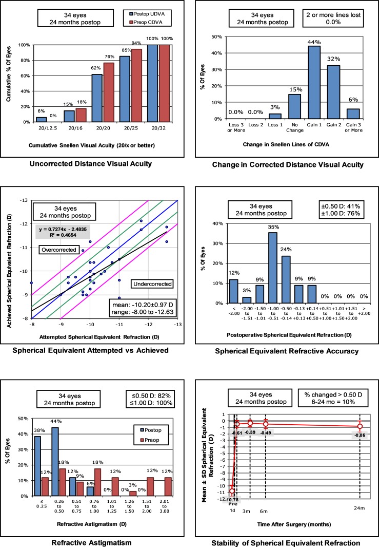
Aim: To investigate the change in posterior corneal elevations (PCEs) of eyes with extremely high myopia 2 years after small incision lenticule extraction (SMILE).

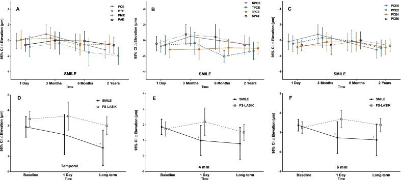
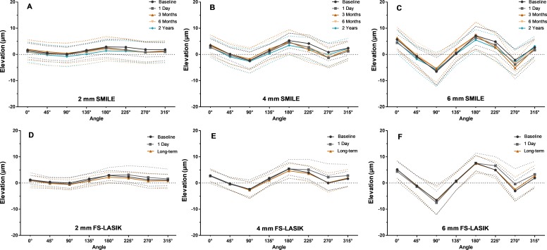
Methods: We evaluated 39 eyes of 39 patients with spherical equivalent higher than -10.00 dioptres (D). Using a Scheimpflug camera (Pentacam), we measured change in PCEs at 1 day, 3 months, 6 months and 2 years after SMILE. Another 34 eyes of 34 patients who underwent femtosecond laser-assisted in situ keratomileusis (FS-LASIK) were examined before, at 1 day and long-term after surgery as the control group. For each eye, elevations at central, thinnest, maximal points and 24 other predetermined points were measured.

Results: No significant forward displacements of PCEs were observed in both surgeries. The maximal but not significant forward displacement occurred around 3-6 months following SMILE, and all returned to original levels 6 months postoperatively except superior area. The peripheral area tended to displace backward, while the central area tended forwardly. In both procedures, elevations along horizontal meridians, inferior and temporal hemispheres were significantly higher than those along vertical meridians, superior and nasal hemispheres, respectively (p<0.05). Elevation on the 4 mm, 6 mm diameters at 1 day and on the 6 mm diameter and temporal hemisphere at long-term follow-up postoperatively were significantly higher in FS-LASIK than SMILE (p<0.05). Change in elevations on the 6 mm diameter circle correlated with residual bed thickness (p=0.047).

Conclusions: SMILE is a safe way to correct for myopia higher than -10 D, with PCEs remaining stable 2 years after surgery.

Keywords: optics and refraction; treatment lasers; treatment surgery.


Conflict of interest statement

Competing interests: None declared.

Figures

Figure 1
Figure 1
Refractive outcomes during the follow-up period after small-incision lenticule extraction (SMILE). CDVA, corrected distance visual acuity;D, dioptres; UDVA, uncorrected distance visual acuity.
Figure 2
Figure 2
Change in posterior corneal elevations after small-incision lenticule extraction (SMILE) and comparison with femtosecond laser-assisted in situ keratomileusis (FS-LASIK). (A) Posterior central (PCE), maximal (PME), posterior average elevation (PAE) and at the thinnest point (PTE). (B) Changes in nasal (NPCE), temporal (TPCE), inferior (IPCE) and superior (SPCE) hemispheres. (C) Changes at centre (PCE0), 2 mm (PCE2), 4 mm (PCE4) and 6 mm (PCE6) diameters. With the exception of △TPCE (p=0.026), no significant changes over time were observed after SMILE. Significant differences between surgeries in TPCE (D), PCE4 (E) and PCE6 (F). Individual points and error bars represent the estimated mean and SD. *P<0.05 between SMILE and FS-LASIK.
Figure 3
Figure 3
Posterior corneal elevations changes at various diameters from the perspective of the meridian after small-incision lenticule extraction (SMILE) and comparison with femtosecond laser-assisted in situ keratomileusis (FS-LASIK). (A–C) Variations at 2, 4 and 6 mm diameter in SMILE group. (D–F) Variations at 2, 4 and 6 mm diameter in FS-LASIK group. Individual points and dotted lines represent the estimated mean and SD.

References

    1. Shah R, Shah S, Sengupta S. Results of small incision lenticule extraction: all-in-one femtosecond laser refractive surgery. J Cataract Refract Surg 2011;37:127–37. 10.1016/j.jcrs.2010.07.033 - DOI - PubMed
    1. Sekundo W, Kunert KS, Blum M. Small incision corneal refractive surgery using the small incision lenticule extraction (SMILE) procedure for the correction of myopia and myopic astigmatism: results of a 6 month prospective study. Br J Ophthalmol 2011;95:335–9. 10.1136/bjo.2009.174284 - DOI - PubMed
    1. Pedersen IB, Ivarsen A, Hjortdal J. Three-year results of small incision Lenticule extraction for high myopia: refractive outcomes and aberrations. J Refract Surg 2015;31:719–24. 10.3928/1081597X-20150923-11 - DOI - PubMed
    1. Wu D, Wang Y, Zhang L, et al. . Corneal biomechanical effects: small-incision lenticule extraction versus femtosecond laser-assisted laser in situ keratomileusis. J Cataract Refract Surg 2014;40:954–62. 10.1016/j.jcrs.2013.07.056 - DOI - PubMed
    1. Belin MW, Khachikian SS. An introduction to understanding elevation-based topography: how elevation data are displayed - a review. Clin Exp Ophthalmol 2009;37:14–29. 10.1111/j.1442-9071.2008.01821.x - DOI - PubMed

Publication types

MeSH terms

LinkOut - more resources