. 2019 May 14;116(20):10019-10024.
doi: 10.1073/pnas.1818544116. Epub 2019 Apr 29.

PGE2 signaling via the neuronal EP2 receptor increases injury in a model of cerebral ischemia

Affiliations

PGE2 signaling via the neuronal EP2 receptor increases injury in a model of cerebral ischemia

Qingkun Liu et al. Proc Natl Acad Sci U S A. .

Abstract

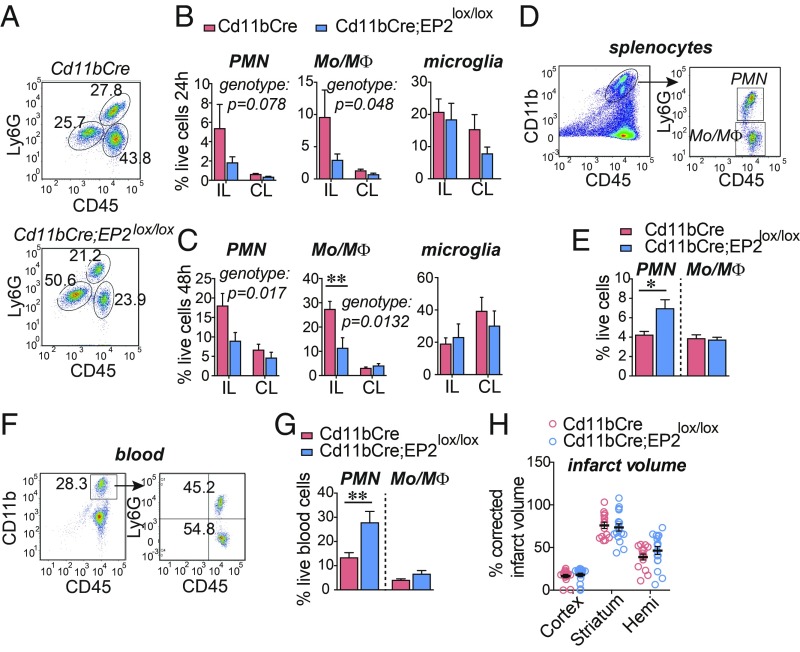
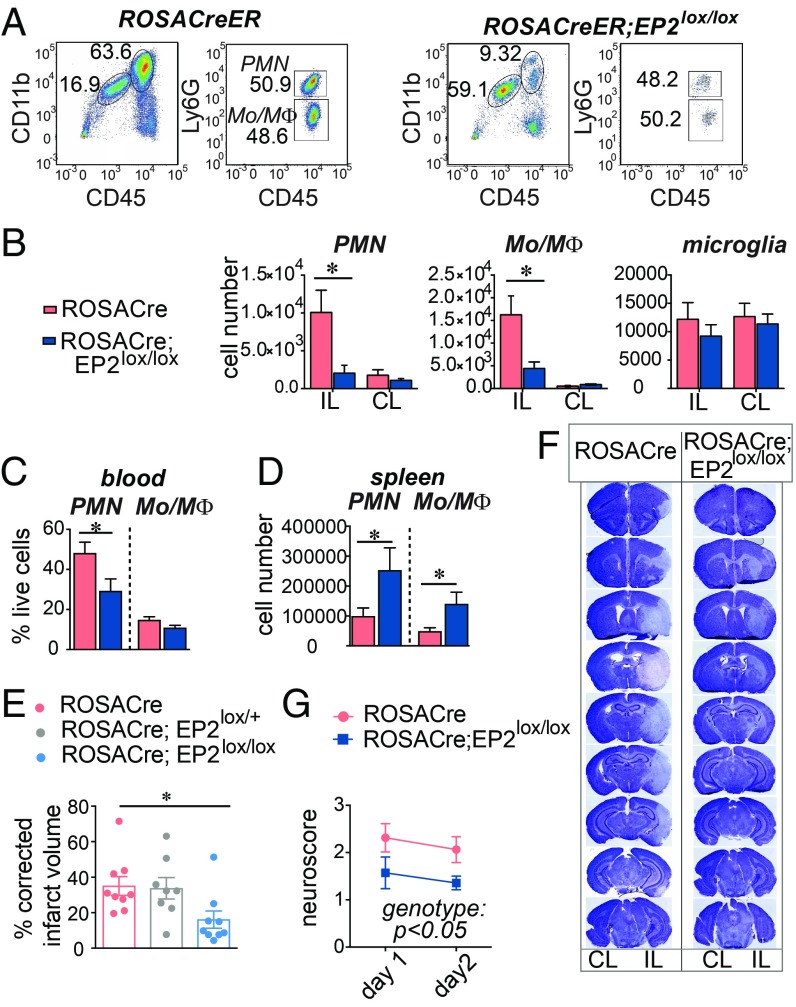
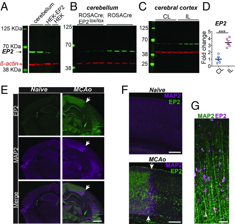
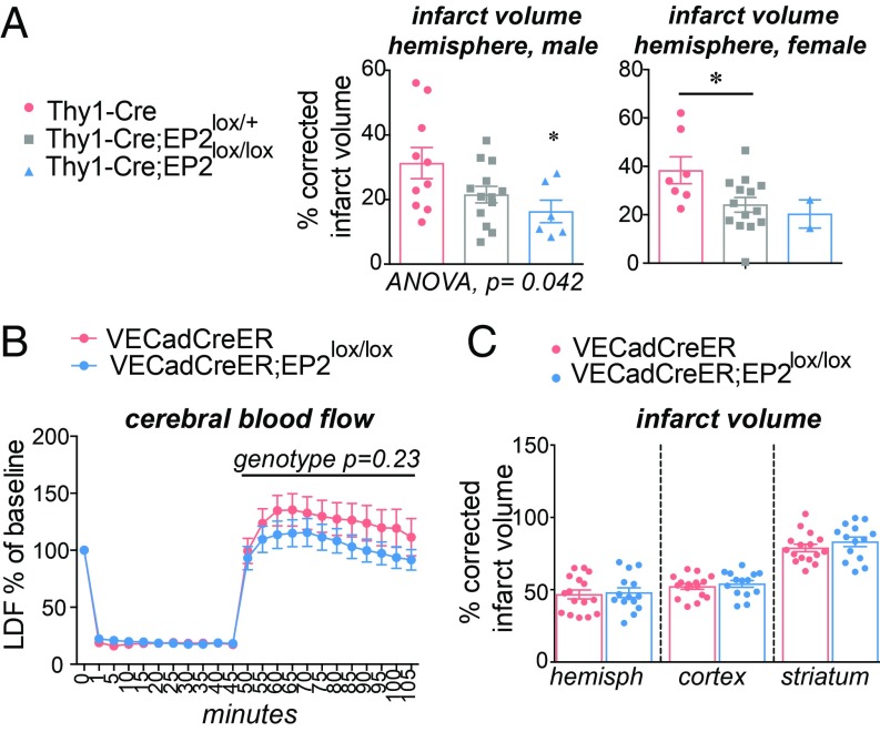
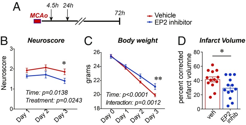
The inflammatory prostaglandin E2 (PGE2) EP2 receptor is a master suppressor of beneficial microglial function, and myeloid EP2 signaling ablation reduces pathology in models of inflammatory neurodegeneration. Here, we investigated the role of PGE2 EP2 signaling in a model of stroke in which the initial cerebral ischemic event is followed by an extended poststroke inflammatory response. Myeloid lineage cell-specific EP2 knockdown in Cd11bCre;EP2lox/lox mice attenuated brain infiltration of Cd11b+CD45hi macrophages and CD45+Ly6Ghi neutrophils, indicating that inflammatory EP2 signaling participates in the poststroke immune response. Inducible global deletion of the EP2 receptor in adult ROSA26-CreERT2 (ROSACreER);EP2lox/lox mice also reduced brain myeloid cell trafficking but additionally reduced stroke severity, suggesting that nonimmune EP2 receptor-expressing cell types contribute to cerebral injury. EP2 receptor expression was highly induced in neurons in the ischemic hemisphere, and postnatal deletion of the neuronal EP2 receptor in Thy1Cre;EP2lox/lox mice reduced cerebral ischemic injury. These findings diverge from previous studies of congenitally null EP2 receptor mice where a global deletion increases cerebral ischemic injury. Moreover, ROSACreER;EP2lox/lox mice, unlike EP2-/- mice, exhibited normal learning and memory, suggesting a confounding effect from congenital EP2 receptor deletion. Taken together with a precedent that inhibition of EP2 signaling is protective in inflammatory neurodegeneration, these data lend support to translational approaches targeting the EP2 receptor to reduce inflammation and neuronal injury that occur after stroke.

Keywords: PGE2; conditional knockout; stroke.


Conflict of interest statement

The authors declare no conflict of interest.

Figures

Fig. 1.
Fig. 1.
Conditional deletion of the EP2 receptor in myeloid cells reduces immune cell infiltration after MCAo. Cd11bCre and Cd11bCre;EP2lox/lox C57B6/J 3 mo male mice underwent MCAo-RP and brain, spleen, and blood myeloid cells were examined. Data are presented as mean ± SEM. (A) Representative plots from Cd11bCre (Top) and Cd11bCre;EP2lox/lox (Bottom) ischemic hemispheres 48 h after MCAo. (B) At 24 h after MCAo, percent live cells in IL and contralateral (CL) hemispheres representing CD11b+CD45hiLy6Ghi neutrophils (PMN), CD11b+CD45hiLy6Glo Mo/MΦ and Cd11b+CD45int microglia are shown (n = 5–8 per group; two-way ANOVA, effects of genotype in italics). (C) At 48 h after MCAo, percentage of live cells representing PMN, Mo/MΦ, and microglia (n = 6 to 7 per group; two-way ANOVA, effects of genotype in italics; post hoc Bonferroni **P < 0.01). (D) Representative gating strategy to identify PMNs and Mo/MΦ in the spleen 48 h after MCAo. (E) At 48 h, percentage of live cells representing PMN and Mo/MΦ in the spleen (n = 8–11 per group; two-tailed Student’s t test, *P < 0.05). (F) Representative gating strategy to identify PMNs and Mo/MΦ in blood 48 h after MCAo. (G) At 48 h, percentage of live cells representing PMN and Mo/MΦ in blood (n = 8–11 per group; two-tailed Student’s t test, **P < 0.01). (H) Percentage of corrected infarct volume for the cortex, striatum, and hemisphere at 72 h after MCAo (n = 15–17 per group).
Fig. 2.
Fig. 2.
Inducible global deletion of the EP2 receptor reduces myeloid cell infiltration after MCAo and prevents cerebral injury. RosaCreER and RosaCreER;EP2lox/lox 3–4 mo C57B6/J male mice underwent MCAo followed by 48 h RP. Data are presented as mean ± SEM. (A) Representative plots of CD11b+CD45hiLy6Ghi PMN and CD11b+CD45hiLy6Glo Mo/MΦ from RosaCreER and RosaCreER;EP2lox/lox ischemic hemispheres 2 d after MCAo. (B) Cell numbers in IL and CL hemispheres for PMN, Mo/MΦ subsets, and microglia (n = 5–11 per group; two-way ANOVA; for PMN, effect of the genotype, P = 0.026, effect of the hemisphere, P = 0.02; for Mo/MΦ, effect of the genotype P = 0.017, effect of the hemisphere, P = 0.002; Tukey post hoc, *P < 0.05). (C) At 48 h, numbers of PMN and Mo/MΦ in the spleen (n = 5–9 per group; two-tailed Student’s t test, *P < 0.05). (D) At 48 h, percentage of live cells representing PMN and Mo/MΦ in blood 48 h after MCAo (n = 7–10 per group; two-tailed Student’s t test, *P < 0.05). (E) Neurological scores (n = 7 to 8 per group; repeated measure two-way ANOVA, effect of the genotype P = 0.0492; effect of time P < 0.0001). (F) Representative series of brain sections stained with Cresyl violet (CV) from RosaCreER and RosaCreER;EP2lox/lox mice 48 h after MCAo. Areas lacking CV staining were quantified for infarct volume. (G) Quantification of the percentage of corrected hemispheric infarct volume at 48 h after MCAo (n = 8 to 9 per group; one-way ANOVA P = 0.0328; Tukey’s post hoc *P < 0.05).
Fig. 3.
Fig. 3.
EP2 receptor is highly induced in the brain in response to cerebral ischemia. (A) Validation of the anti-EP2 antibody using cerebellar lysates and HEK cells overexpressing the EP2 receptor (HEK-EP2; positive control) and HEK cell lysates (negative control). (B) EP2 receptor expression is decreased in the cerebellum in ROSACreER;EP2lox/lox 3 mo male mice. (C) The EP2 receptor is significantly induced in the IL cerebral cortex compared with the CL noninfarcted cortex 48 h after MCAo in 3 mo male mice. (D) Quantification of the EP2 protein in the IL and CL cortices 48 h after MCAo (n = 5 per group, two-tailed Student’s t test, ***P = 0.0003). Data are presented as mean ± SEM. (E) Immunofluorescent staining of naive and MCAo brains 48 h after MCAo, stained for the EP2 receptor and MAP2. (Scale bar, 1 mm.) The white arrow points to the border among infarcted tissue and penumbra. (F) Higher magnification of the border region between the infarcted and the penumbral parietal cortex and the corresponding area in the naive brain. (Scale bar, 250 μm.) (G) Layers V and VI of the ischemic hemisphere in the penumbra demonstrating colocalization of MAP2 (green) and the EP2 receptor (purple). (Scale bar, 50 μm.).
Fig. 4.
Fig. 4.
Effects of cell-specific deletion of the neuronal and endothelial EP2 receptors in MCAo-RP. Data are presented as mean ± SEM. (A) Deletion of the neuronal EP2 receptor in 3 to 4 mo Thy1-Cre;EP2lox/lox mice was protective in male and female cohorts (males: n = 6–10/group; P = 0.0417; females: n = 2–14/group; P = 0.0470; *P < 0.05 post hoc Tukey between male Thy1-Cre and Thy1-Cre;EP2lox/lox genotypes). (B) Measurement of relative CBF by LDF does not demonstrate any effect of genotype in VECadCreER;EP2lox/lox vs. control VECadCreER mice (n = 14–20 3 to 4 mo male mice per group; effect of genotype during RP from 45 min to 105 min, P = 0.2365). (C) Percentage of corrected infarct volume does not differ in 3 to 4 mo male VECadCreER;EP2lox/lox mice vs. control VECadCreER mice 24 h after MCAo (n = 14–16 per group).
Fig. 5.
Fig. 5.
Inhibition of the EP2 receptor is cerebroprotective. Data are presented as mean ± SEM. (A) The EP2 receptor inhibitor (compound 52 at 10 mg/kg) or vehicle were administered at 4.5 h and again at 24 h after MCAo to 3 mo C57BL/6J male mice (n = 21 per group). (B) Neurological scores over 72 h after MCAo (repeated measures, two-way ANOVA, effect of time P = 0.0138; effect of treatment, P = 0.0243; Bonferroni post hoc *P < 0.05 at day 3). (C) Body weight over 72 h after MCAo (repeated measures, two-way ANOVA, effect of time P < 0.0001, and effect of treatment, P = 0.0052; Tukey post hoc **P < 0.01 at day 3). (D) Percentage of infarct volume in mice 72 h after MCAo ± EP2 receptor inhibitor (n = 13 per group; Student’s two-tailed t test, P = 0.035).

Similar articles

Cited by

References

    1. Kawano T, et al. (2006) Prostaglandin E2 EP1 receptors: Downstream effectors of COX-2 neurotoxicity. Nat Med 12:225–229. - PubMed
    1. Liang X, et al. (2011) Signaling via the prostaglandin E₂ receptor EP4 exerts neuronal and vascular protection in a mouse model of cerebral ischemia. J Clin Invest 121:4362–4371. - PMC - PubMed
    1. Shi J, et al. (2010) The prostaglandin E2 E-prostanoid 4 receptor exerts anti-inflammatory effects in brain innate immunity. J Immunol 184:7207–7218. - PMC - PubMed
    1. Johansson JU, et al. (2015) Prostaglandin signaling suppresses beneficial microglial function in Alzheimer’s disease models. J Clin Invest 125:350–364. - PMC - PubMed
    1. Liang X, et al. (2005) Deletion of the prostaglandin E2 EP2 receptor reduces oxidative damage and amyloid burden in a model of Alzheimer’s disease. J Neurosci 25:10180–10187. - PMC - PubMed

Publication types

Substances