Case Reports
. 2019 May 14;116(20):9865-9870.
doi: 10.1073/pnas.1817815116. Epub 2019 Apr 29.

Mutations in MAGT1 lead to a glycosylation disorder with a variable phenotype

Affiliations
Case Reports

Mutations in MAGT1 lead to a glycosylation disorder with a variable phenotype

Eline Blommaert et al. Proc Natl Acad Sci U S A. .

Abstract

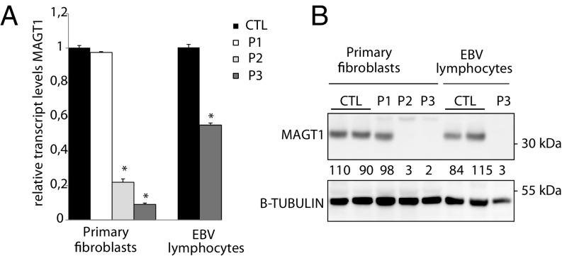
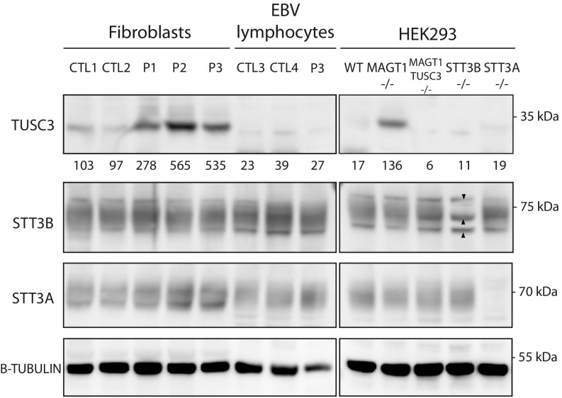
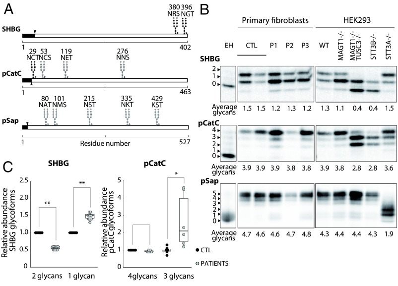
Congenital disorders of glycosylation (CDG) are a group of rare metabolic diseases, due to impaired protein and lipid glycosylation. We identified two patients with defective serum transferrin glycosylation and mutations in the MAGT1 gene. These patients present with a phenotype that is mainly characterized by intellectual and developmental disability. MAGT1 has been described to be a subunit of the oligosaccharyltransferase (OST) complex and more specifically of the STT3B complex. However, it was also claimed that MAGT1 is a magnesium (Mg2+) transporter. So far, patients with mutations in MAGT1 were linked to a primary immunodeficiency, characterized by chronic EBV infections attributed to a Mg2+ homeostasis defect (XMEN). We compared the clinical and cellular phenotype of our two patients to that of an XMEN patient that we recently identified. All three patients have an N-glycosylation defect, as was shown by the study of different substrates, such as GLUT1 and SHBG, demonstrating that the posttranslational glycosylation carried out by the STT3B complex is dysfunctional in all three patients. Moreover, MAGT1 deficiency is associated with an enhanced expression of TUSC3, the homolog protein of MAGT1, pointing toward a compensatory mechanism. Hence, we delineate MAGT1-CDG as a disorder associated with two different clinical phenotypes caused by defects in glycosylation.

Keywords: CDG; XMEN; congenital disorders of glycosylation; oligosaccharyltransferase complex.


Conflict of interest statement

The authors declare no conflict of interest.

Figures

Fig. 1.
Fig. 1.
Expression levels of MAGT1. (A) Relative transcript levels of MAGT1 and (B) protein immunoblotting for MAGT1 in fibroblasts and EBV-transformed lymphocytes. β-Tubulin was used as a loading control. Values below the corresponding lane represent averaged normalized values (n = 3). Error bars represent SE of mean (SEM). *P < 0.05.
Fig. 2.
Fig. 2.
Stability of different OST subunits. Fibroblasts, EBV-transformed lymphocytes, and HEK293 cells were analyzed for protein steady state levels of TUSC3, STT3B, and STT3A. The arrowheads depict the nonspecific bands comigrating with STT3B. β-Tubulin was used as a loading control. Values below the TUSC3 blot represent averaged values normalized to the average of the control fibroblast cells (n = 3).
Fig. 3.
Fig. 3.
STT3B-dependent glycosylation is affected in patients’ fibroblasts. (A) Diagrams showing the glycosylation sites of SHBG, pCatC, and pSap. Black glycan structures indicate an STT3B-dependent site. Signal sequences are depicted in black. (B) HEK293 cells and fibroblasts were transfected with SHBG, pCatC, or pSap, followed by pulse-chase labeling. Quantified values are shown below gel lanes and represent the average number of glycans for the respective reporter (n = 3). (C) Quantification of the different glycoforms of SHBG and pCatC in fibroblasts, normalized to the averaged control samples. EH indicates endoglycosidase H treatment. *P < 0.5; **P < 0.005.
Fig. 4.
Fig. 4.
Glycosylation assessment of different substrates. (A) Assessment of SHBG glycosylation. MAGT1−/− TUSC3−/− cells were cotransfected with SHBG and the indicated MAGT1 transcripts. (B) Metabolic labeling of the endogenous pCatC and GLUT1 in HEK293 cells and EBV-transformed lymphocytes. (C) HEK293 cells, primary fibroblasts, and EBV-transformed lymphocytes were analyzed for protein steady state levels of pCatC and CatC. β-Tubulin was used as a loading control. (D) Diagram showing the STT3B-dependent glycosylation site of GLUT1. The signal sequence is depicted in black. (E) Relative abundance of the 4- and 3-glycans form for pCatC. (F) Relative steady state levels of pCatC and CatC in primary fibroblasts and EBV-transformed lymphoctyes. CatC was not quantifiable in HEK293 cells. Quantified values are shown below gel lanes and represent the average number of glycans for the respective reporter (n = 3). EH indicates endoglycosidase H treatment. Error bars represent SEM. *P < 0.05.

References

    1. Jaeken J, Péanne R. What is new in CDG? J Inherit Metab Dis. 2017;40:569–586. - PubMed
    1. Freeze HH, Chong JX, Bamshad MJ, Ng BG. Solving glycosylation disorders: Fundamental approaches reveal complicated pathways. Am J Hum Genet. 2014;94:161–175. - PMC - PubMed
    1. Aebi M. N-linked protein glycosylation in the ER. Biochim Biophys Acta Mol Cell Res. 2013;1833:2430–2437. - PubMed
    1. Kelleher DJ, Karaoglu D, Mandon EC, Gilmore R. Oligosaccharyltransferase isoforms that contain different catalytic STT3 subunits have distinct enzymatic properties. Mol Cell. 2003;12:101–111. - PubMed
    1. Ruiz-Canada C, Kelleher DJ, Gilmore R. Cotranslational and posttranslational N-glycosylation of polypeptides by distinct mammalian OST isoforms. Cell. 2009;136:272–283. - PMC - PubMed

Publication types

MeSH terms

LinkOut - more resources