. 2019 Jun 21;201(14):e00724-18.
doi: 10.1128/JB.00724-18. Print 2019 Jul 15.

A Genome-Wide Helicobacter pylori Morphology Screen Uncovers a Membrane-Spanning Helical Cell Shape Complex

Affiliations

A Genome-Wide Helicobacter pylori Morphology Screen Uncovers a Membrane-Spanning Helical Cell Shape Complex

Desirée C Yang et al. J Bacteriol. .

Abstract

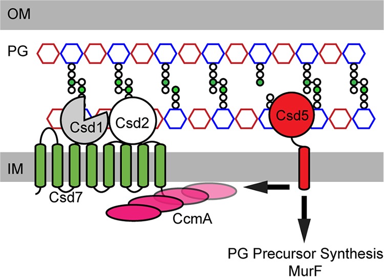
Evident in its name, the gastric pathogen Helicobacter pylori has a helical cell morphology which facilitates efficient colonization of the human stomach. An improved light-focusing strategy allowed us to robustly distinguish even subtle perturbations of H. pylori cell morphology by deviations in light-scattering properties measured by flow cytometry. Profiling of an arrayed genome-wide deletion library identified 28 genes that influence different aspects of cell shape, including properties of the helix, cell length or width, cell filament formation, cell shape heterogeneity, and cell branching. Included in this mutant collection were two that failed to form any helical cells, a soluble lytic transglycosylase and a previously uncharacterized putative multipass inner membrane protein HPG27_0728, renamed Csd7. A combination of cell fractionation, mutational, and immunoprecipitation experiments show that Csd7 and Csd2 collaborate to stabilize the Csd1 peptidoglycan (PG) endopeptidase. Thus, both csd2 and csd7 mutants show the same enhancement of PG tetra-pentapeptide cross-linking as csd1 mutants. Csd7 also links Csd1 with the bactofilin CcmA via protein-protein interactions. Although Csd1 is stable in ccmA mutants, these mutants show altered PG tetra-pentapeptide cross-linking, suggesting that Csd7 may directly or indirectly activate as well as stabilize Csd1. These data begin to illuminate a highly orchestrated program to regulate PG modifications that promote helical shape, which includes nine nonessential nonredundant genes required for helical shape and 26 additional genes that further modify H. pylori's cell morphology.IMPORTANCE The stomach ulcer and cancer-causing pathogen Helicobacter pylori has a helical cell shape which facilitates stomach infection. Using light scattering to measure perturbations of cell morphology, we identified 28 genes that influence different aspects of cell shape. A mutant in a previously uncharacterized protein renamed Csd7 failed to form any helical cells. Biochemical analyses showed that Csd7 collaborates with other proteins to stabilize the cell wall-degrading enzyme Csd1. Csd7 also links Csd1 with a putative filament-forming protein via protein-protein interactions. These data suggest that helical cell shape arises from a highly orchestrated program to regulate cell wall modifications. Targeting of this helical cell shape-promoting program could offer new ways to block infectivity of this important human pathogen.

Keywords: Helicobacter pylori; cell shape; flow cytometry; peptidoglycan; stomach infection.


Figures

FIG 1
FIG 1
Light scattering profiles distinguish diverse shape phenotypes in an ordered H. pylori deletion library. (A to I) Forward versus side scatter contour plots measured by flow cytometry showing the wild-type and the indicated mutant populations. Drawn gates represent 64% of the wild-type population. For cell shape mutants, the numbers listed refer to the percentages of the populations in this wild-type gate. Insets, light micrographs (×1,000 magnification, phase-contrast) of strains with indicated shapes. For the contour plots: blue and green represent areas of lower cell density, red and orange correspond to areas of high cell density, and yellow is midrange. Scale bars, 2 μm. Strains used were wild type (WT; LSH100), ccmA (LSH142), csd3 (NSH152a), slt (MHH9), HPG27_1093 (DCY8a), HPG27_1325 (DCY113), prc (HPG27_1298; DCY114), HPG27_0119 (DCY115), and HPG27_0750 (DCY6a).
FIG 2
FIG 2
csd7 and slt are essential for normal helical shape. Complementation strains have the native locus disrupted by deletion and insertion of chloramphenicol acetyltransferase cassette (cat) and the indicated gene expressed from the rdxA locus. (A) Light micrographs (×1,000 magnification, phase-contrast) of various strains (genotypes listed below). Scale bars, 5 μm. (B) Forward versus side scatter contour plot measured by flow cytometry showing the csd7 mutant population. The drawn red gate encompasses 64% of the control wild-type population. For csd7, 48% of the mutant population falls within this control gate. (C) Scatter plot arraying wild-type, csd7, and the csd7 complemented (csd7 rdxA::csd7) populations by cell length (x axis) versus side curvature (y axis). Quantitative analyses of phase-contrast images of bacteria to measure side curvature and central axis length were performed with the CellTool software package as described previously (9). (D) Smooth histogram displaying population cell curvature (x axis) as function of density (y axis). (E) Scatter plot arraying wild-type, slt, and the slt complemented (slt rdxA::slt) populations by cell length (x axis) versus cell curvature (y axis). (F) Smooth histogram displaying population cell curvature (x axis) as function of density (y axis). Strains used were WT (LSH100), slt (MHH9), slt rdxA::slt (AC1), csd7:cat rdxA::csd7 (DCY28), and csd7:cat (DCY7a).
FIG 3
FIG 3
Csd7 is an integral membrane protein. (A) Schematic of the csd7 gene locus identified in the flow cytometry screen. The disruption site of the chloramphenicol acetyltransferase cassette (cat) was inserted 347 bp in the same orientation as the HPG27_0728 open reading frame and results in a 60-bp internal deletion. (B) Predicted topology for Csd7. IM, inner membrane. (C) Cell fractionation and localization of Csd7-FLAG with and without detergent (0.5% Triton X-100). The whole-cell extract (WCE), pellet (PEL), and supernatant (SUP) fractions were analyzed by SDS-PAGE and immunoblotted for Csd7-FLAG using anti-FLAG antibodies. The molecular weight marker (kDa) is on the left. Strains used: LSH100 and DCY71.
FIG 4
FIG 4
Csd1, Csd2, and Csd7 show costability dependency, and catalytically inactive variants of Csd1 and Csd2 do not affect protein stability. Whole-cell extracts of the indicated strains, normalized by OD600, were analyzed by SDS-PAGE and immunoblotted with anti-Csd1 (A, E, and F), anti-FLAG (B and D), anti-CcmA (C), or anti-VSV-G (G) antibodies. Positions of the molecular weight markers (in kilodaltons) are on the left. Complemented strains are indicated with “C.” LS, unmarked LSH100 strain; csd2-F, Csd2-FLAG; csd7-F, Csd7-FLAG; csd2-V, Csd2-VSV-G strain. The gene listed above each band indicates the relevant deletion or mutation made at the native loci. Strains used: ACH1, DCY7a, DCY28, DCY72, DCY73, DCY74, DCY75, DCY76, DCY77, DCY89, DCY102, DCY106, DCY105, DCY110, DCY111, DCY112, JTH4, LSH100, LSH113, LSH120, LSH121, LSH140, LSH141, LSH142, LSH120, and NAH1.
FIG 5
FIG 5
Csd1, Csd2, and Csd7 directly interact with each other in bacterial two hybrid (BATCH) and co-immunoprecipitation (co-IP) assay blots. (A to D) Plasmid pairs encoding indicated T18 and T25 fusion proteins were cotransformed into E. coli BTH101 (cya-99). Individual colonies were patched on M9-glucose supplemented with Amp, Kan, X-Gal, and 1 mM IPTG. Plates were incubated at room temperature and photographed after 72 h. Interacting partners bring together T18 and T25 to reconstitute adenylate cyclase activity that is detected using lacZ induction (blue) as a reporter. (E) FLAG co-IP of Csd1-FLAG and Western blotted for FLAG-tagged Csd1 and VSV-G-tagged Csd2. (F) FLAG co-IP of Csd2-FLAG and Western blotted for FLAG-tagged Csd2 and Csd1. (G) VSV-G co-IP of Csd7-VSV-G and Western blotted for VSV-G-tagged Csd7 and FLAG-tagged Csd2. (H) Protein stability codependencies. The arrows represent promotion of protein stability, black dashed lines indicate protein interactions validated by pulldown under native conditions, and the gray dashed line indicates a putative protein interaction validated by pulldown using formaldehyde cross-linking.
FIG 6
FIG 6
csd7 alters cell morphology in strain PMSS1. (A) Scatter plot arraying wild-type, csd1, csd7, and the csd7 complemented (csd7 rdxA::csd7) populations by cell length (x axis) versus side curvature (y axis). Quantitative analysis of phase-contrast images of bacteria to measure side curvature and central axis length was performed with the CellTool software package as described previously (9). (B) Smooth histogram displaying population cell curvature (x axis) as function of density (y axis). Strains used: DCY119, DCY120, LMH1, and PMSS1.
FIG 7
FIG 7
csd7 promotes stomach colonization. Stomach colonization loads from 1-week infections of csd7 mutant (Δcsd7), complemented (cmpl), and wild-type strains in pairwise competition as indicated in the MSD132 (left) and PMSS1 (right) strain backgrounds. Each data point represents the CFU per gram of stomach tissue for each genotype from a single mouse. Lines connect the data points of each genotype from one animal and the dotted line indicates the limit of detection of infection. Numbers below the x axis indicate the fractions of mice infected with each genotype. Displayed data were pooled from two separate experiments, and each replicate is shown in black or gray. Differences in colonization loads among genotype in competition were assessed by Wilcoxon matched pairs signed-rank test. *, P < 0.05; **, P < 0.01. Strains used: DCY27, DCY119, DCY120, DCY121, MSD132, and PMSS1.
FIG 8
FIG 8
Model of the Csd7 protein interaction network. Shown is a schematic diagram of a putative “helical shapesome” which includes the PG hydrolase Csd1, nonenzymatic proteins (CcmA, Csd2, Csd7, and Csd5) that serve to stabilize and/or localize this complex, and the PG precursor synthesis enzyme MurF. See the text for details. OM, outer membrane; IM, inner membrane; PG, peptidoglycan.

References

    1. Kusters JG, van Vliet AHM, Kuipers EJ. 2006. Pathogenesis of Helicobacter pylori infection. Clin Microbiol Rev 19:449–490. doi:10.1128/CMR.00054-05. - DOI - PMC - PubMed
    1. Ferlay J, Soerjomataram I, Dikshit R, Eser S, Mathers C, Rebelo M, Parkin DM, Forman D, Bray F. 2015. Cancer incidence and mortality worldwide: sources, methods and major patterns in GLOBOCAN 2012. Int J Cancer 136:E359–E386. doi:10.1002/ijc.29210. - DOI - PubMed
    1. Goodwin CS, McCulloch RK, Armstrong JA, Wee SH. 1985. Unusual cellular fatty acids and distinctive ultrastructure in a new spiral bacterium (Campylobacter pyloridis) from the human gastric mucosa. J Med Microbiol 19:257–267. doi:10.1099/00222615-19-2-257. - DOI - PubMed
    1. Montecucco C, Rappuoli R. 2001. Living dangerously: how Helicobacter pylori survives in the human stomach. Nat Rev Mol Cell Biol 2:457–466. doi:10.1038/35073084. - DOI - PubMed
    1. Cover TL, Blaser MJ. 2009. Helicobacter pylori in health and disease. Gastroenterology 136:1863–1873. doi:10.1053/j.gastro.2009.01.073. - DOI - PMC - PubMed

Publication types

LinkOut - more resources