. 2019 Jul;74(7):675-683.
doi: 10.1136/thoraxjnl-2018-212529. Epub 2019 Apr 29.

Urban airborne particle exposure impairs human lung and blood Mycobacterium tuberculosis immunity

Affiliations

Urban airborne particle exposure impairs human lung and blood Mycobacterium tuberculosis immunity

Martha Torres et al. Thorax. 2019 Jul.

Abstract

Rationale: Associations between urban (outdoor) airborne particulate matter (PM) exposure and TB and potential biological mechanisms are poorly explored.

Objectives: To examine whether in vivo exposure to urban outdoor PM in Mexico City and in vitro exposure to urban outdoor PM2.5 (< 2.5 µm median aerodynamic diameter) alters human host immune cell responses to Mycobacterium tuberculosis.

Methods: Cellular toxicity (flow cytometry, proliferation assay (MTS assay)), M. tuberculosis and PM2.5 phagocytosis (microscopy), cytokine-producing cells (Enzyme-linked immune absorbent spot (ELISPOT)), and signalling pathway markers (western blot) were examined in bronchoalveolar cells (BAC) and peripheral blood mononuclear cells (PBMC) from healthy, non-smoking, residents of Mexico City (n=35; 13 female, 22 male). In vivo-acquired PM burden in alveolar macrophages (AM) was measured by digital image analysis.

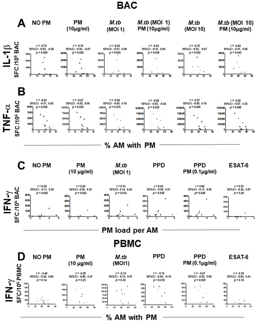
Measurements and main results: In vitro exposure of AM to PM2.5 did not affect M. tuberculosis phagocytosis. High in vivo-acquired AM PM burden reduced constitutive, M. tuberculosis and PM-induced interleukin-1β production in freshly isolated BAC but not in autologous PBMC while it reduced constitutive production of tumour necrosis factor-alpha in both BAC and PBMC. Further, PM burden was positively correlated with constitutive, PM, M. tuberculosis and purified protein derivative (PPD)-induced interferon gamma (IFN-γ) in BAC, and negatively correlated with PPD-induced IFN-γ in PBMC.

Conclusions: Inhalation exposure to urban air pollution PM impairs important components of the protective human lung and systemic immune response against M. tuberculosis. PM load in AM is correlated with altered M. tuberculosis-induced cytokine production in the lung and systemic compartments. Chronic PM exposure with high constitutive expression of proinflammatory cytokines results in relative cellular unresponsiveness.

Keywords: alveolar macrophages; particulate matter; pollution; tuberculosis.


Conflict of interest statement

Competing interests: None declared.

Figures

Figure 1
Figure 1
Cytotoxic effects of PM2.5 are observed only at high PM2.5 concentrations. BAC and PBMC (each n=6) were exposed to 0, 0.1, 1, 10 and 25 μg/mL of PM2.5 and incubated for 24–72 hours to evaluate particulate matter (PM) exposure effects on cell viability (A–D) and induction of apoptosis (E–H). (A) Proportions of live alveolar macrophages (AM), (B) peripheral blood monocytes (PBMN), (C) alveolar lymphocytes (AL), and (D) peripheral blood lymphocytes (PBL) were examined by a LIVE/DEAD Fixable Far Red Dead Cell Stain and analysed by flow cytometry with fluorescence detection at 660 nm (A–D). Apoptosis was identified with a far red-emitting DNA-intercalating dye by flow cytometry using a 488 nm laser with fluorescence detection at 570 nm (Apoptosis Detection Reagent) in a flourescence-activated cell sorting (FACS) fusion cytometer. (E) AM, (F) PBMN, (G) AL and (H) PBL. Friedman and Dunn’s post hoc test for multiple comparison was performed. The p values above horizontal lines represent the indicated significant differences comparison. BAC, bronchoalveolar cell; PBMC, peripheral blood mononuclear cell.
Figure 2
Figure 2
Assessment of PM burden in AM. Wright-stained cytospin preparations of bronchoalveolar cells (BAC) (120 000 BACs per cytospin) from 30 participants (CC #) were examined to assess the proportions of AM containing PM (%AM with PM) (A) and the mean and minimum and maximum areas of the total AM area occupied by PM (PM load per AM) (B) in each study participant. Digital colour images of 50 alveolar macrophages per study participant were obtained using 1000× magnified digital bright field microscopy (Olympus BX51 microscope, Olympus, Waltham, MA). PM load per AM (areas of AM occupied by PM) was determined using ImageJ V.1.49. The areas occupied by PM within each of the AMs are expressed as per cent of the total (100%) AM area. The grey bars represent the study participants whose BACs were assessed by enzyme-linked immune absorbent spot (ELISPOT) (see figures 3–5). AM, alveolar macrophage; PM, particulate matter.
Figure 3
Figure 3
Proinflammatory cytokine production in BAC and peripheral blood mononuclear cells (PBMC) grouped by in vivo-acquired PM burden of AM. Frequencies of constitutive BAC-spot-forming cells (A) and frequencies of IL-1β (B), TNF-α (C) and IL-6 (D) spot-forming cells in PM exposed and/ or Mycobacterium tuberculosis infected, BAC (from n=9 participants) evaluated by enzyme-linked immune absorbent spot (ELISPOT) assay. Closed red symbols represent participants with ≥44.3% AM with PM and open symbols represent participants with <42.6% AM with PM (range: high proportion 44.3%–60%, low proportion 26%–42.6%). Mann-Whitney test was performed using the GraphPad Prism software for Mac OS X V.6.0. P values above symbols represent significant differences in frequencies of cytokine-producing cells between groups with higher and lower %AM with PM for the corresponding experimental condition. For example, in panel B, frequencies of IL-1β-producing BAC following M. tuberculosis infection (MOI1) and PM2.5 exposure from study participants with higher %AM with PM (red symbols) are compared with BAC following M. tuberculosis infection (MOI1) and PM2.5 exposure from study participants with lower %AM with PM (open symbols). AM, alveolar macrophage; BAC, bronchoalveolar cell; IFN, interferon; IL, interleukin; PM, particulate matter; TNF, tumour necrosis factor.
Figure 4
Figure 4
Proinflammatory cytokine production in BAC and PBMC grouped by in vivo-acquired PM burden of AM. (A) Frequencies of IFN-γ-spot-forming cells in PM exposed and/or Mycobacterium tuberculosis infected, PPD and 6 kDa early secretory antigenic target (ESAT-6)-stimulated BAC (from nine participants). (B) Frequencies of IFN-γ-spot-forming cells in PM exposed and/or M. tuberculosis infected, PPD and ESAT-6-stimulated PBMC evaluated by enzyme-linked immune absorbent spot (ELISPOT) assay. Closed blue symbols (A) represent high (≥2.24%) in vivo inhalation-acquired PM load (PM load per AM) and open symbols represent participants with low PM load per AM (<2.24%). Closed red symbols (B) represent participants with ≥44.3% AM with PM and open symbols represent participants with <42.6% AM with PM (range: high proportion 44.3%–60%, low proportion 26%–42.6%). Mann-Whitney test was performed using the GraphPad Prism software for Mac OS X V.6.0. P values above symbols represent significant differences in frequencies of cytokine-producing cells between groups with higher and lower PM load per AM (blue symbols) (A) or higher and lower %AM% with PM (red symbols) (B) for the corresponding experimental condition. For example, in panel A, frequencies of IFN-γ-producing BAC following M. tuberculosis infection (MOI1) and PM2.5 exposure from individuals with higher in vivo inhalation-acquired PM load per AM (blue symbols) were compared with BAC from individuals with lower in vivo inhalation-acquired PM load per AM (open symbols). AM, alveolar macrophages; BAC, bronchoalveolar cells; IFN, interferon; PBMC, peripheral blood mononuclear cells; PM, particulate matter; PPD, purified protein derivative.
Figure 5
Figure 5
Correlations between in vivo-acquired PM burden in AM (%AM with PM or PM load per AM) and frequencies of cytokine-producing BAC and PBMC. Frequencies of IL-1β (A), TNF-α (B) and IFN-γ (C) spot-forming cells (SFC) in BAC (n=9) and frequencies of IFN-γ-SFC in PBMC (n=9) (D) are shown as a function of PM burden in AM following exposure to PM2.5 and/or M.tuberculosis infection, or PPD and ESAT-6 stimulation. Data points represent participants and slopes’ positive or negative correlations. Spearman’s r correlation coefficient test was performed using the GraphPad Prism software for Mac OS X V.6.0. A CI for all statistics test of 95% (α=0.05) was used. The p values, r values and CIs are indicated in all the experimental conditions. AM, alveolar macrophages; BAC, bronchoalveolar cells; IFN, interferon; IL, interleukin; M.tuberculosis, Mycobacterium tuberculosis; PBMC, peripheral blood mononuclear cells; PM, particulate matter; PPD, purified protein derivative; TNF, tumour necrosis factor; ESAT-6, 6kDa secretory antigenic target.

References

    1. Nations U. World urbanization prospects: the 2018 revision, 2018.
    1. Organization WH. Who fact sheet No. 292, household air pollution and health 2016. Available: http://www.who.int/mediacentre/factsheets/fs292/en/
    1. Bourdrel T, Bind M-A, Béjot Y, et al. Cardiovascular effects of air pollution. Arch Cardiovasc Dis 2017;110:634–42. - PMC - PubMed
    1. Bowe B, Xie Y, Li T, et al. Global and national burden of diabetes mellitus attributable to PM2.5 air pollution. Lancet Planet Health 2016;2018:e301–12. - PubMed
    1. Saenen ND, Bové H, Steuwe C, et al. Children’s urinary environmental carbon load. A novel marker reflecting residential ambient air pollution exposure? Am J Respir Crit Care Med 2017;196:873–81. - PubMed

Publication types

MeSH terms