. 2019 Oct;40(10):1334-1342.
doi: 10.1038/s41401-019-0233-9. Epub 2019 Apr 29.

Identification of PRDX6 as a regulator of ferroptosis

Affiliations

Identification of PRDX6 as a regulator of ferroptosis

Bin Lu et al. Acta Pharmacol Sin. 2019 Oct.

Abstract

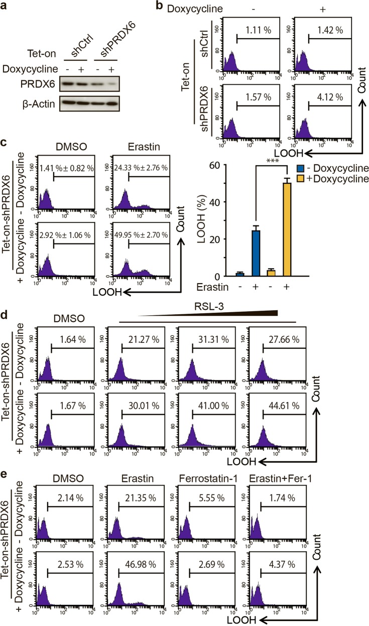
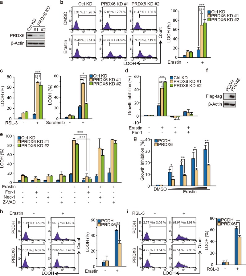
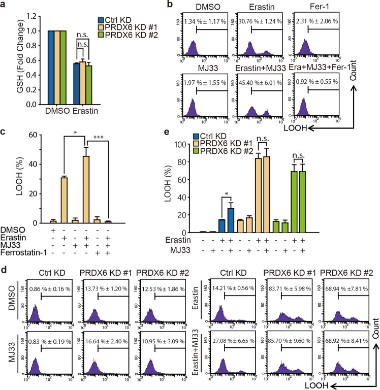
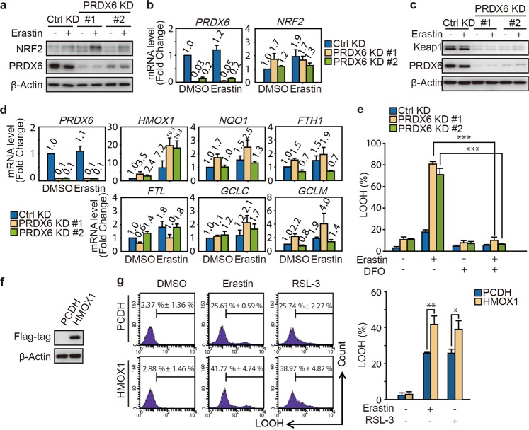
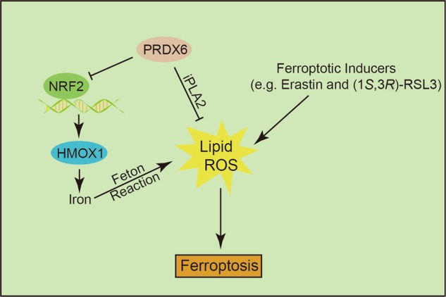
Ferroptosis is a newly characterized iron-dependent form of nonapoptotic regulated cell death triggered by lipid reactive oxygen species (LOOH). The dysregulation of ferroptosis is highly related to cancer, and the induction of ferroptosis is also proposed as a potential strategy for cancer therapy. Although several key regulators have been identified that are involved in ferroptosis, the molecular mechanism underlying this process remains largely unknown. Here, we report that Peroxiredoxin-6 (PRDX6) is a bona fide negative regulator of ferroptotic cell death. The knockdown of intracellular PRDX6 significantly enhances LOOH and ferroptotic cell death triggered by ferroptosis inducers (Erastin and RSL-3), which is correlated with the transcriptional activation of heme oxygenase-1. Moreover, overexpression of heme oxygenase-1 enhances both Erastin- and RSL-3-triggered LOOH, suggesting that heme oxygenase-1 mediates PRDX6 silencing-enhanced ferroptosis. More importantly, the application of a specific PRDX6 phospholipase A2 (iPLA2) inhibitor, MJ-33, synergistically enhances the ferroptosis induced by Erastin, suggesting that PRDX6 removes LOOH through its iPLA2 activity. Thus, our findings reveal an essential role of PRDX6 in protecting cells against ferroptosis and provide a potential target to improve the antitumor activity of ferroptosis-based chemotherapy.

Keywords: PRDX6; antitumor therapy; ferroptosis; heme oxygenase-1; iPLA2 activity.


Conflict of interest statement

The authors declare no competing interests.

Figures

Fig. 1
Fig. 1
PRDX6 knockdown through inducible lentiviral knockdown system enhances ferroptotic LOOH. a Western blot analysis of PRDX6 expression in indicated genetically engineered H1299 cells after doxycycline treatment (0.25 μg/mL) for 48 h, b LOOH production was assayed by flow cytometry using C11-BODIPY. c After doxycycline (0.25 μg/mL) treatment for 48 h, indicated genetically engineered H1299 cells were treated with Erastin (2 μM) for 12 h, and LOOH production was assayed. Representative data are shown and the statistical data are from three independent experiments and the bar indicates the mean value (***P < 0.001). d After doxycycline treatment (0.25 μg/mL) for 48 h, indicated genetically engineered H1299 cells were treated with RSL-3 (50, 100, or 150 nM) for 12 h, and LOOH production was assayed. e After doxycycline (0.25 μg/mL) treatment for 48 h, indicated genetically engineered H1299 cells were treated with Erastin (2 μM) with or without ferrostatin-1 (10 μM) for 12 h, and LOOH production was assayed
Fig. 2
Fig. 2
PRDX6 knockdown through transient knockdown system enhances ferroptotic cell death. a Western blot analysis of PRDX6 expression in H1299 after infection of lentivirus targeting to PRDX6 for 48 h. b After infection for 48 h, H1299 cells was treated with Erastin (2 μM) for 12 h, and LOOH production was assayed by flow cytometry using C11-BODIPY. Representative data are shown and the statistical analysis is from nine independent experiments and the bar indicates the mean values. c After infection for 48 h, H1299 cells were treated with RSL-3 (50 nM) or sorafenib (20 μM) for 12 h, and LOOH production was assayed by flow cytometry using C11-BODIPY. The statistical analysis is from three independent experiments and the bar indicates the mean values. d After infection for 48 h, H1299 cells were treated for 24 h with Erastin (2 μM) with or without ferrostatin-1(Fer-1) (10 μM), and cell viability was assayed by SRB assay. The statistical analysis is from three independent experiments and the bar indicates the mean values. e After infection for 48 h, H1299 cells were treated with Erastin (2 μM) with or without Fer-1 (10 μM), necrostatin-1 (30 μM) and Z-VAD-FMK (10 μM) for 12 h, and LOOH production was assayed. The statistical analysis is from three independent experiments and the bar indicates the mean values. f Western blot analysis of PRDX6 overexpression in H1299 cells with transfection of PRDX6 cDNA. g After gene transfection for 24 h, H1299 cells are treated with Erastin (0, 1, 2, 4, and 8 μM) for 24 h, and cell viability was assayed by SRB assay. The statistical analysis is from three independent experiments and the bar indicates the mean values. h, i After gene transfection for 24 h, H1299 cells are treated with Erastin (8 μM) or RSL-3 (100 nM) for 12 h, and LOOH production was assayed. Representative data are shown and the statistical analysis is from three independent experiments and the bar indicates the mean values. (*P < 0.05; **P < 0.01; ***P < 0.001)
Fig. 3
Fig. 3
PRDX6 negatively regulates ferroptosis through its iPLA2 activity. a After infection of lentivirus targeting to PRDX6 for 48 h, H1299 cells were treated with Erastin (2 μM) for 6 h and intracellular GSH levels were examined. The statistical analysis is from three independent experiments and the bar indicates the mean values. b, c H1299 cells were treated with Erastin (2 μM) with or without of MJ33(5 μM) or ferrostatin-1 (10 μM) for 12 h, and LOOH production was assayed by flow cytometry using C11-BODIPY. Representative data are shown and the statistical analysis is from three independent experiments and the bar indicates the mean values. d, e After infection for 48 h, H1299 cells were treated with Erastin (2 μM) with or without MJ33 (5 μM) for 12 h, and LOOH production was assayed. Representative data are shown and the statistical analysis is from three independent experiments and the bar indicates the mean values. (*P < 0.05; ***P < 0.001; n.s.: no statistic difference)
Fig. 4
Fig. 4
PRDX6 silencing promotes the transcriptional activation of heme oxygenase-1. a, b After infection of lentivirus targeting to PRDX6 for 48 h, H1299 cells were treated with Erastin (2 μM) for 12 h. a The protein expression of NRF2, and b its mRNA expression were assayed. The relative gene expression is normalized to β-actin and the error bar indicates the s.d. value from triplicates. c After infection for 48 h, H1299 cells were treated with Erastin (2 μM) for 12 h, and Western blot analysis of Keap1 and PRDX6 expression. d After infection for 48 h, H1299 cells were treated with Erastin (2 μM) for 12 h. These NRF2 downstream genes were assayed. The relative gene expression is normalized to β-actin and the error bar indicates the s.d. value from triplicates. e After infection for 48 h, H1299 cells were treated with Erastin (2 μM) with or without DFO (100 μM) for 12 h, and LOOH production was assayed. Representative data are shown and the statistical analysis is from three independent experiments and the bar indicates the mean values. f Western blot analysis of HMOX1 overexpression in H1299 cells with transfection of  HMOX-1 cDNA. g After gene transfection for 24 h, H1299 cells are treated with Erastin (2 μM) or RSL-3 (50 nM) for 12 h, and LOOH production was assayed. Representative data are shown and the statistical analysis is from three independent experiments and the bar indicates the mean values. (*P < 0.05; **P < 0.01; ***P < 0.001)
Fig. 5
Fig. 5
The schematic representation of PRDX6 negatively regulating ferroptosis working model

References

    1. Dixon SJ, Lemberg KM, Lamprecht MR, Skouta R, Zaitsev EM, Gleason CE, et al. Ferroptosis: an iron-dependent form of nonapoptotic cell death. Cell. 2012;149:1060–72. doi: 10.1016/j.cell.2012.03.042. - DOI - PMC - PubMed
    1. Yang WS, Kim KJ, Gaschler MM, Patel M, Shchepinov MS, Stockwell BR. Peroxidation of polyunsaturated fatty acids by lipoxygenases drives ferroptosis. Proc Natl Acad Sci USA. 2016;113:E4966–75. doi: 10.1073/pnas.1603244113. - DOI - PMC - PubMed
    1. Doll S, Proneth B, Tyurina YY, Panzilius E, Kobayashi S, Ingold I, et al. ACSL4 dictates ferroptosis sensitivity by shaping cellular lipid composition. Nat Chem Biol. 2017;13:91–8. doi: 10.1038/nchembio.2239. - DOI - PMC - PubMed
    1. Kagan VE, Mao G, Qu F, Angeli JP, Doll S, Croix CS, et al. Oxidized arachidonic and adrenic PEs navigate cells to ferroptosis. Nat Chem Biol. 2017;13:81–90. doi: 10.1038/nchembio.2238. - DOI - PMC - PubMed
    1. Stockwell BR, Friedmann Angeli JP, Bayir H, Bush AI, Conrad M, Dixon SJ, et al. Ferroptosis: a regulated cell death nexus linking metabolism, redox biology, and disease. Cell. 2017;171:273–85. doi: 10.1016/j.cell.2017.09.021. - DOI - PMC - PubMed