. 2019 Jul;4(7):1088-1095.
doi: 10.1038/s41564-019-0432-7. Epub 2019 Apr 29.

Ectosymbiotic bacteria at the origin of magnetoreception in a marine protist

Affiliations

Ectosymbiotic bacteria at the origin of magnetoreception in a marine protist

Caroline L Monteil et al. Nat Microbiol. 2019 Jul.

Abstract

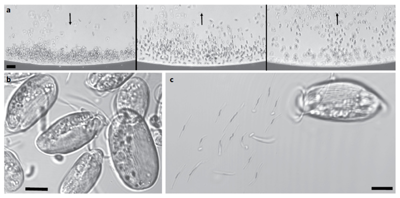
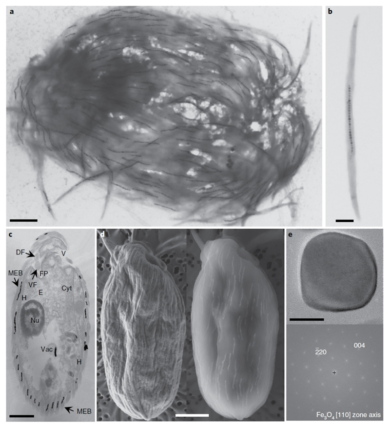
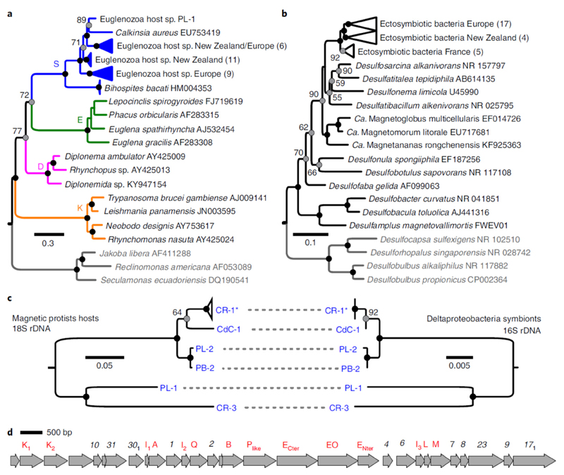
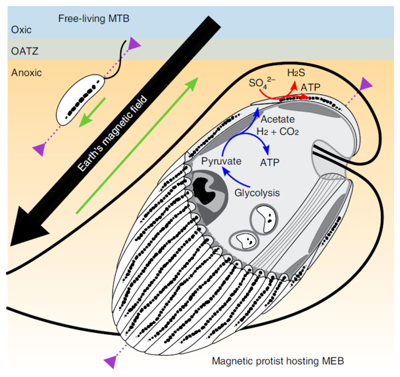
Mutualistic symbioses are often a source of evolutionary innovation and drivers of biological diversification1. Widely distributed in the microbial world, particularly in anoxic settings2,3, they often rely on metabolic exchanges and syntrophy2,4. Here, we report a mutualistic symbiosis observed in marine anoxic sediments between excavate protists (Symbiontida, Euglenozoa)5 and ectosymbiotic Deltaproteobacteria biomineralizing ferrimagnetic nanoparticles. Light and electron microscopy observations as well as genomic data support a multi-layered mutualism based on collective magnetotactic motility with division of labour and interspecies hydrogen-transfer-based syntrophy6. The guided motility of the consortia along the geomagnetic field is allowed by the magnetic moment of the non-motile ectosymbiotic bacteria combined with the protist motor activity, which is a unique example of eukaryotic magnetoreception7 acquired by symbiosis. The nearly complete deltaproteobacterial genome assembled from a single consortium contains a full magnetosome gene set8, but shows signs of reduction, with the probable loss of flagellar genes. Based on the metabolic gene content, the ectosymbiotic bacteria are anaerobic sulfate-reducing chemolithoautotrophs that likely reduce sulfate with hydrogen produced by hydrogenosome-like organelles6 underlying the plasma membrane of the protist. In addition to being necessary hydrogen sinks, ectosymbionts may provide organics to the protist by diffusion and predation, as shown by magnetosome-containing digestive vacuoles. Phylogenetic analyses of 16S and 18S ribosomal RNA genes from magnetotactic consortia in marine sediments across the Northern and Southern hemispheres indicate a host-ectosymbiont specificity and co-evolution. This suggests a historical acquisition of magnetoreception by a euglenozoan ancestor from Deltaproteobacteria followed by subsequent diversification. It also supports the cosmopolitan nature of this type of symbiosis in marine anoxic sediments.


Conflict of interest statement

Competing interests

The authors declare no competing interests.

Figures

Fig. 1
Fig. 1
Light microscope images of south-seeking magnetic protists sampled in the Mediterranean Sea, Carry-le-Rouet, France. a, The microscope was focused on a point at the edge of the water droplet closest to the north pole of the bar magnet, producing a local field direction indicated by the black arrow (left). Reversing the bar magnet so that the south magnetic pole was closest to the edge of the drop caused south-seeking organisms to rotate and swim in the opposite direction towards the opposite edge of the droplet (middle and right, also indicated by black arrows). b,c, Confocal images of the striated, magnetically responsive protists (b) and their disaggregation 20 min after deposition between a slide and coverslip, showing the presence of rod-shaped bacteria detached from the surface of the protist (c). Scale bars, 20 μm (a) and 5 μm (b,c).
Fig. 2
Fig. 2
Electron microscopy images of the magnetic protist sampled in the Mediterranean Sea, Carry-le-Rouet. a,b, TEM images showing the ultrastructure of a single magnetic consortium containing about 150 magnetosome chains (a) and a magnetic ectosymbiotic bacterium detached from its host (b). c, TEM image of the longitudinal section through a single magnetic consortium showing the general morphological features of the magnetic protist, such as the nucleus (Nu), a battery of extrusomes (E), the vestibulum (V), the cytostome (Cyt), MEB on the extracellular matrix, the flagellar pocket (FP) with the dorsal and ventral flagella (DF and VF, respectively), hydrogenosomes (H) or mitochondria-like organelles in close vicinity to the ectosymbionts, and digestive vacuoles (Vac) in which grazed magnetotactic bacteria and their magnetosomes can be seen. d, Images of a single magnetic consortium observed using a SEM operating at 2 kV (left) or 10 kV (right) showing the presence of magnetosome chains in the bacteria that cover the protist. e, High-resolution TEM image of a single magnetosome biomineralized by an ectosymbiotic bacterium (top) and the corresponding fast Fourier transform (bottom) for which labelled reflexions have been indexed with respect to the magnetite structure. No octahedral or elongated asymmetric shapes are clearly visible (Supplementary Fig. 3). Scale bars, 2 μm (a,c,d), 0.5 μm (b) and 20 nm (e).
Fig. 3
Fig. 3
Diversity of the magnetic protists and their ectosymbionts. a,b, Phylogenetic trees based on 18S rRNA and 16S rRNA gene sequences showing the evolutionary relationships of the magnetic protists with the Euglenozoa (a; S, Symbiontida; E, Euglenida; D, Diplonemida and K, Kinetoplastida) and the MEB with the Desulfobacteraceae (b), respectively. The trees were rooted with other Excavata (protists) and Desulfobacterales (bacteria) families (species in grey). The number of clones obtained are indicated in parenthesis. The numbers next to the grey circles at nodes represent the proportional bootstrap support values. The black circles represent nodes supported by 100% of the replicates. The GenBank accession numbers are also shown. c, Host and symbiont phylogenies built from a subsample of sequences obtained from individual single holobionts only. Topology congruence provides evidence of co-evolution. CR-1* represents a collapsed clade of four single holobionts for which the 16S sequences were identical. CR, Carry-le-Rouet; PL, Port Leucate; PB, Port de Boulouris and CdC, Cap de Creus. d, Chromosomal section containing a magnetosome gene cluster showing the organization of different mam (red) and mad (black) genes of the MEB from a representative holobiont CR-1 isolated from Carry-le-Rouet.
Fig. 4
Fig. 4
Schematic illustration of the magnetotactic consortium showing the magnetotactic behaviour in the Northern Hemisphere and the syntrophic interactions between partners. The green arrows show the anterior–posterior orientation of the organisms, which is parallel and antiparallel to the Earth’s magnetic field lines for the freeliving magnetotactic bacteria (MTB) and the consortium protist-MEB, respectively. The purple arrows show the organism’s motility zone in the sediments. The ATP synthesis by the hydrogenosomes in the protist is symbolized by the blue arrows. Molecular hydrogen, acetate and carbon dioxide are products that could be transported through the plasma and used by the MEB as sources of energy and carbon. The red arrows show the dissimilatory sulfate reduction by MEB, which uses hydrogen as an electron donor and produces hydrogen sulfide outside the consortium. OATZ, oxic–anoxic transition zone.

Comment in

  • Symbiotic magnetic motility.
    Edgcomb V. Edgcomb V. Nat Microbiol. 2019 Jul;4(7):1066-1067. doi: 10.1038/s41564-019-0489-3. Nat Microbiol. 2019. PMID: 31222171 No abstract available.

References

    1. Margulis L, Fester R. Symbiosis as a Source of Evolutionary Innovation: Speciation and Morphogenesis. MIT Press; 1991. - PubMed
    1. López-García P, Eme L, Moreira D. Symbiosis in eukaryotic evolution. J Theor Biol. 2017;434:20–33. - PMC - PubMed
    1. Bernhard JM, Buck KR, Farmer MA, Bowser SS. The Santa Barbara Basin is a symbiosis oasis. Nature. 2000;403:77–80. - PubMed
    1. Morris BEL, Henneberger R, Huber H, Moissl-Eichinger C. Microbial syntrophy: interaction for the common good. FEMS Microbiol Rev. 2013;37:384–406. - PubMed
    1. Yubuki N, Leander BS. Diversity and evolutionary history of the symbiontida (Euglenozoa) Front Ecol Evol. 2018;6:100.

Publication types

MeSH terms