. 2019 Apr 15:10:374.
doi: 10.3389/fneur.2019.00374. eCollection 2019.

Corticospinal Tract Integrity and Long-Term Hand Function Prognosis in Patients With Stroke

Affiliations

Corticospinal Tract Integrity and Long-Term Hand Function Prognosis in Patients With Stroke

Yeun Jie Yoo et al. Front Neurol. .

Abstract

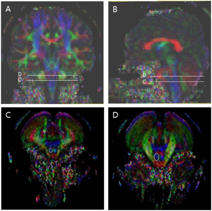
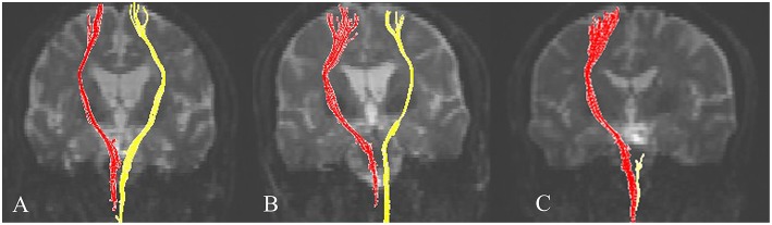
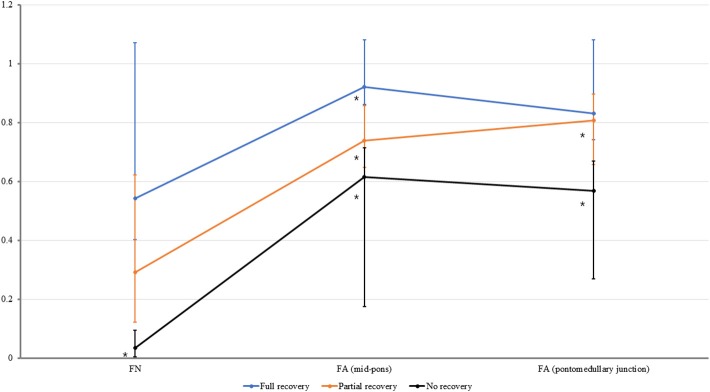
Background: The restoration of hand function is an important goal for patients with stroke. This study investigated the relationship between corticospinal tract (CST) integrity and the functional status of the hand in patients with stroke 6 months after onset and evaluated which of the following values would be useful for predicting hand function: fiber number (FN), fractional anisotropy (FA) at the mid-pons, and FA at the pontomedullary junction. Methods: The present retrospective cross-sectional observational study assessed 44 patients with stroke who were able to walk without using a walking aid or orthosis. The final hand function results were classified into three groups: no recovery (unable to grasp), partial recovery (able to grasp, unable to oppose), and full recovery (able to grasp and oppose). All subjects underwent diffusion tensor imaging (DTI) at 6 months after stroke onset. Values for FA at the mid-pons and pontomedullary junction and CST FN were measured. The normalization ratio for FN and FA was calculated using the following formula: data for affected hand/data for non-affected hand. Results: The normalized FN, FA (mid-pons), and FA (pontomedullary junction) DTI values differed significantly. The FA (mid-pons) value for the full recovery group was higher than those for the other groups. The FA (mid-pons) value for the partial recovery group was higher than that for the no recovery group. The normalized FA (mid-pons) value differed significantly among all three groups. Conclusions: The present study showed that CST integrity (at 6 months after onset) in patients with chronic stroke was related to functional hand status. In addition, the mid-pons FA value was more predictive of functional restoration of the hand than the FN or FA value at the pontomedullary junction. These results may be useful in predicting the functional restoration of the hand and understanding the functional prognosis of stroke.

Keywords: corticospinal tract; diffusion tensor imaging; hand function; prognosis; stroke; stroke rehabilitation.


Figures

Figure 1
Figure 1
Regions of interest (ROIs) used to reconstruct the corticospinal tract (CST) on diffusion tensor imaging. The stippled lines indicate the ROIs. The solid lines indicate the seed ROI for the corticospinal tract. (A) Coronal color FA map; (B) Sagittal color FA map; (C) The seed ROI of CST at the pontomedullary junction; (D) The seed ROI of CST at the mid-pons.
Figure 2
Figure 2
Representative diffusion tensor tractography images of the corticospinal tract in typical subjects from the three groups. Subjects from the (A) full recovery, (B) partial recovery, and (C) no recovery groups. The non-affected tract is shown in red, and the affected tract in yellow.
Figure 3
Figure 3
The normalized fiber number (FN), fractional anisotropy (FA) at the mid-pons, and FA at the pontomedullary junction of the corticospinal tract in all groups. The median values are shown as dots. For all values, the error bar shows the interquartile range between the first and third quartiles. *The FN value for no recovery group was lower than those that for full recovery group (P ≤ 0.0166). *The FA (mid-pons) value for the full recovery group was higher than those for the other two groups (P ≤ 0.0166), and the FA (mid-pons) value for the partial recovery group was higher than that for the no recovery group (P ≤ 0.0166). *The FA (pontomedullary junction) value for the full recovery group and partial recovery group were higher than that for the no recovery group (P ≤ 0.0166, respectively). The FA (pontomedullary junction) value for the full recovery group and that for partial recovery group were not significantly different.

Similar articles

Cited by

References

    1. Jorgensen HS, Nakayama H, Raaschou HO, Vive-Larsen J, Stoier M, Olsen TS. Outcome and time course of recovery in stroke. Part II: time course of recovery the Copenhagen stroke study. Arch Phys Med Rehabil. (1995) 76:406–12. 10.1016/S0003-9993(95)80568-0 - DOI - PubMed
    1. Chen CL, Tang FT, Chen HC, Chung CY, Wong MK. Brain lesion size and location: effects on motor recovery and functional outcome in stroke patients. Arch Phys Med Rehabil. (2000) 81:447–52. 10.1053/mr.2000.3837 - DOI - PubMed
    1. Binkofski F, Seitz RJ, Hacklander T, Pawelec D, Mau J, Freund HJ. Recovery of motor functions following hemiparetic stroke: a clinical and magnetic resonance-morphometric study. Cerebrovasc Dis. (2001) 11:273–81. 10.1159/000047650 - DOI - PubMed
    1. Shelton FN, Reding MJ. Effect of lesion location on upper limb motor recovery after stroke. Stroke. (2001) 32:107–12. 10.1161/01.STR.32.1.107 - DOI - PubMed
    1. Lee KB, Kim JS, Hong BY, Kim YD, Hwang BY, Lim SH. The motor recovery related with brain lesion in patients with intracranial hemorrhage. Behav Neurol. (2015) 2015:258161. 10.1155/2015/258161 - DOI - PMC - PubMed