

Role of multi-parametric ultrasound in transient perivascular inflammation of the carotid artery syndrome
- PMID: 31037091
- PMCID: PMC6475971
- DOI: 10.1177/1742271X18822658
Role of multi-parametric ultrasound in transient perivascular inflammation of the carotid artery syndrome
Abstract
Introduction: The term "carotidynia" has been used to describe a symptom or a nosologic entity characterized by pain in the lateral neck region and over the carotid bifurcation. Recent advances in diagnostic imaging and the introduction of diagnostic criteria have led to the adoption of term "Transient perivascular inflammation of the carotid artery" (TIPIC) syndrome.
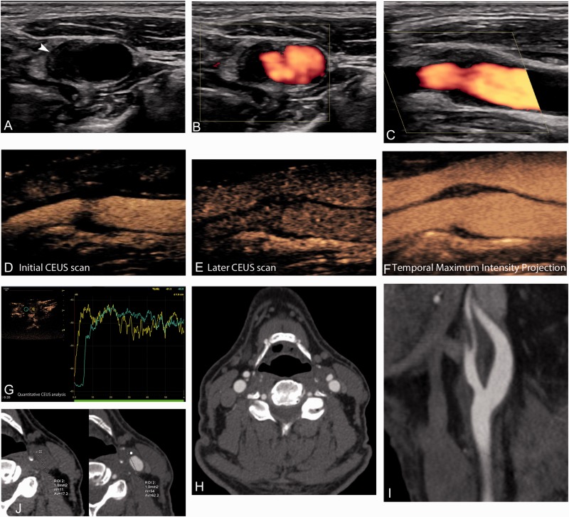
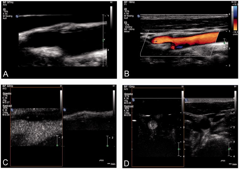
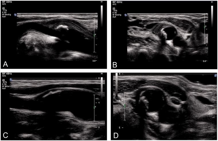
Method: A retrospective analysis of the Radiology Department's database was performed to identify cases with the diagnosis of TIPIC syndrome. The purpose was to identify ultrasound images including B-mode technique, colour, power Doppler technique and contrast-enhanced ultrasound (CEUS).
Findings: In total, five patients with the diagnosis of TIPIC syndrome are presented in this review. TIPIC syndrome is a clinic-radiologic entity characterized by pain over the carotid area, a symptom referring to a wide differential diagnosis where imaging plays a crucial role for proper diagnosis and treatment. Characteristic imaging findings on conventional ultrasound and CEUS are presented in this review.
Discussion: TIPIC syndrome can be investigated with virtually any imaging modality. Ultrasound typically reveals perivascular infiltration and a hypoechoic intimal plaque, while no significant luminal narrowing is appreciated. Computed tomography angiography and magnetic resonance angiography also demonstrate these vascular wall changes primarily affecting the distal common carotid artery, the carotid bulb and possibly the internal carotid artery proximal part. Contrast enhancement is a very characteristic and constant finding of TIPIC lesions, suggestive of the inflammatory nature of the disease and can be appreciated on computed tomography angiography and magnetic resonance angiography. CEUS has been recently used and successfully observed contrast enhancement of the lesions, similar to computed tomography angiography and magnetic resonance angiography.
Conclusion: Ultrasound remains the first-line modality for the evaluation of TIPIC syndrome, capable of providing all the information needed, especially if supplemented with the administration of microbubbles so that the enhancement of lesions can be evaluated.
Keywords: Carotid; contrast-enhanced ultrasound; transient perivascular inflammation of the carotid artery syndrome; ultrasound.
Figures
References
-
- Headache Classification Subcommittee of the International Headache Society. The International Classification of Headache Disorders. 2nd edition. Cephalalgia 2004; 24(Suppl 1): 9–160. - PubMed
-
- Headache Classification Committee of the International Headache Society. Classification and diagnostic criteria for headache disorders, cranial neuralgias and facial pain.. Cephalalgia 1988; 8(Suppl 7): 1–96. - PubMed
-
- Stanbro M, Gray BH, Kellicut DC. Carotidynia: revisiting an unfamiliar entity. Ann Vasc Surg 2011; 25: 1144–1153. - PubMed
Publication types
LinkOut - more resources
Full Text Sources