Review
. 2019 May;104(5):517-529.
doi: 10.1007/s00223-019-00550-z. Epub 2019 Apr 29.

Fibrous Dysplasia of Bone and McCune-Albright Syndrome: A Bench to Bedside Review

Affiliations
Review

Fibrous Dysplasia of Bone and McCune-Albright Syndrome: A Bench to Bedside Review

Iris Hartley et al. Calcif Tissue Int. 2019 May.

Abstract

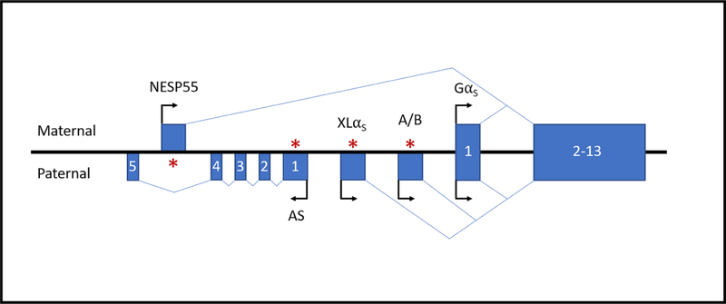
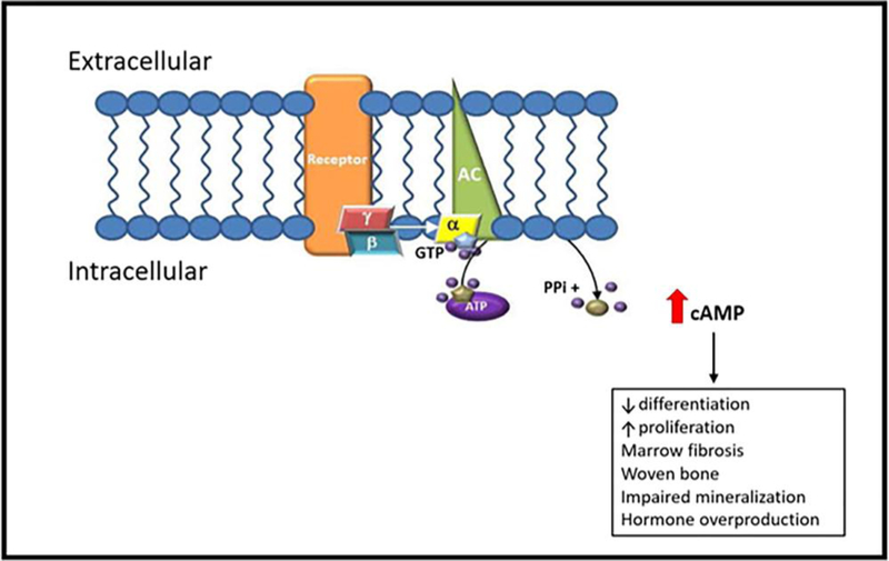
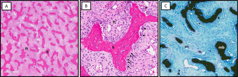
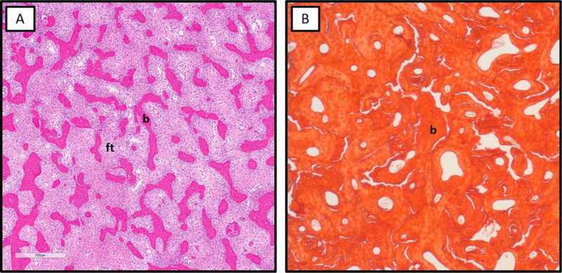
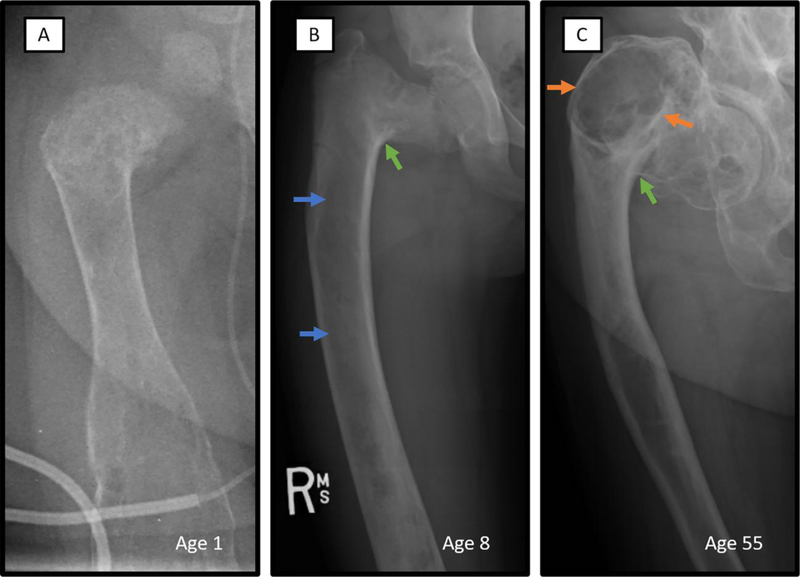
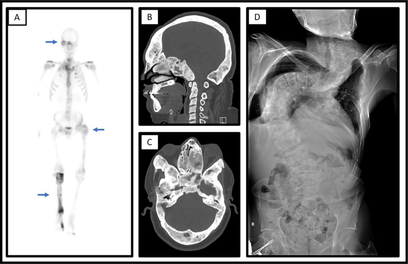
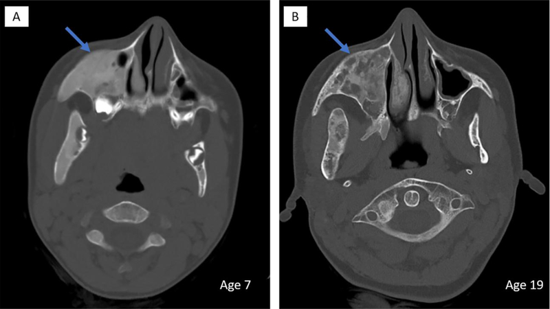
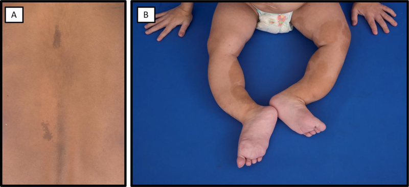
Fibrous dysplasia is an uncommon mosaic disorder in which bone is replaced by structurally unsound fibro-osseous tissue. It is caused by the sporadic post-zygotic activating mutations in GNAS, resulting in dysregulated GαS-protein signaling in affected tissues. This manifests on a broad clinical spectrum ranging from insignificant solitary lesions to severe disease with deformities, fractures, functional impairment, and pain. Fibrous dysplasia may present in isolation or in association with hyperfunctioning endocrinopathies and café-au-lait macules, known as McCune-Albright Syndrome. This review summarizes the current understanding of pathophysiology in fibrous dysplasia, describes key pre-clinical and clinical investigations, and details the current approach to diagnosis and management.

Keywords: Bone disorders; Bone metabolism; FGF23-mediated hypophosphatemia; McCune–Albright syndrome.


Figures

Figure 1.
Figure 1.. Schematic representation of the GNAS locus on chromosome 20q13.3.
The GNAS locus encodes four primary sense transcripts, GαS, XLαS,NESP55, and A/B, and an antisense transcript GNAS-AS1. The sense transcripts are generated by alternative splicing of four unique first exons to common exons 2–13. Allelic and tissue-dependent expression of the XLαS, NESP55, A/B, and GNAS-AS1 transcripts is regulated by differential methylation of their promoters. The GαS transcript exhibits random asymmetric bi-allelic expression in bone. Boxes and connecting lines represent the exons and introns, respectively. Arrows indicate the direction of transcription. Asterisks indicate methylation of the imprinted promoters on either the maternal or paternal allele. Figure is not drawn to scale.
Figure 2.
Figure 2.. G protein-coupled signaling upregulation in FD/MAS.
Fibrous dysplasia is caused by post-zygotic mutations in GNAS, which result in the expression of a constitutively signaling GαS protein. The mutant GαS activates adenylyl cyclase in a ligand-independent manner, leading to excess production of cyclic AMP and protracted downstream signaling. In bone, this promotes the proliferation of immature osteoblast progenitor cells, which leads to formation of structurally abnormal matrix, with increased fibrosis, decreased mineralization, cortical thinning, and obliteration of hematopoietic bone marrow. AC, adenylyl cyclase; α/β/γ, alpha/beta/gamma-subunits of Gs; GTP, guanosine triphosphate; ATP, adenosine triphosphate; PPi, pyrophosphate; cAMP, cyclic adenosine monophosphate. Adapted from Burke AB, et al, Oral Dis. 2017 Sep; 23(6): 697–708.
Figure 3.
Figure 3.. Characteristic histologic features of fibrous dysplasia.
A. Sharpey’s fibers (collagen fibers oriented perpendicular to the bone surface, arrows) and abundant osteoclasts (arrowheads) at the surface of FD bone trabeculae. B. Von Kassa staining showing undermineralization of FD bone with excess osteoid (os) and paucity of mineralized bone matrix (mb). C. Osteogenic cells on the surface of bone trabeculae have a retracted “stellate” appearance (arrows).
Figure 4.
Figure 4.. Histologic patterns of fibrous dysplasia.
A. Typical pattern of FD bone with thin discontinuous trabeculae (b) surrounded by abundant fibrous tissue (ft). B. Sclerosing/pagetoid pattern of FD bone with thick, interconnected trabeculae consisting of woven bone and relative paucity of fibrous tissue.
Figure 5.
Figure 5.. Radiographic features of fibrous dysplasia.
Typical X-ray features of FD, including age-related progression, is demonstrated in three different patients with extensive involvement of the right femur. A. Radiograph of the right femur of a 1-year-old with has the heterogeneous appearance commonly seen in infants with FD. B. Prototypical FD lesion of an 8-year-old demonstrates homogeneous ‘ground glass’ radiolucency (blue arrows) and severe shepherd’s crook deformity (green arrow). C. Sclerotic-appearing FD lesion of older adulthood is shown in the right femur radiograph of a 57-year-old. Typical ‘rind’ sign consisting of a radiolucent lesion surrounded by sclerotic bone (orange arrows) and a severe shepherd’s crook deformity (green arrow) are shown.
Figure 6.
Figure 6.. Characteristic imaging findings in fibrous dysplasia.
A. Numerous areas of increased uptake on Technetium-MDP bone scan (blue arrows) consistent with polyostotic fibrous dysplasia in an adult man. B/C. Computed tomography scan shows extensive involvement (blue arrows) of the skull and facial bones in a 28-year-old male with craniofacial FD, with diffuse areas of expansile bone “ground glass” appearing bone. Shown are sagittal (B) and transverse (C) views. D. Radiographs show severe scoliosis in a 36-year-old man with polyostotic fibrous dysplasia/McCune Albright syndrome.
Figure 7.
Figure 7.. Age-related radiographic changes in craniofacial fibrous dysplasia.
Head CT scans of the same patient at age 7 (A) and age 19 (B) demonstrates typical age-related radiographic changes in the appearance of craniofacial FD. Areas of homogeneous appearing abnormal bone in the right maxilla adopts a more heterogeneous appearance with radiolucent regions with increased age (blue arrows).
Figure 8.
Figure 8.. Typical café-au-lait skin macules in McCune-Albright syndrome.
A. An 8-year-old girl has small café-au-lait macules reflecting along the midline of the back. B) A 2-year-old girl has extensive café-au-lait macules on her bilateral lower extremities. Note the bowing deformities resulting from severe fibrous dysplasia.

References

    1. El-Mofty SK, Fibro-osseous lesions of the craniofacial skeleton: an update. Head Neck Pathol, 2014. 8(4): p. 432–44. - PMC - PubMed
    1. Yang L, et al., Prevalence of Different Forms and Involved Bones of Craniofacial Fibrous Dysplasia. J Craniofac Surg, 2017. 28(1): p. 21–25. - PubMed
    1. Weinstein LS, et al., Activating mutations of the stimulatory G protein in the McCune-Albright syndrome. N Engl J Med, 1991. 325(24): p. 1688–95. - PubMed
    1. Michienzi S, et al., GNAS transcripts in skeletal progenitors: evidence for random asymmetric allelic expression of Gs alpha. Hum Mol Genet, 2007. 16(16): p. 1921–30. - PubMed
    1. Hilger D, Masureel M, and Kobilka BK, Structure and dynamics of GPCR signaling complexes. Nat Struct Mol Biol, 2018. 25(1): p. 4–12. - PMC - PubMed

MeSH terms

LinkOut - more resources