

Imaging in Retinopathy of Prematurity
- PMID: 31037876
- PMCID: PMC7891847
- DOI: 10.22608/APO.201963
Imaging in Retinopathy of Prematurity
Abstract
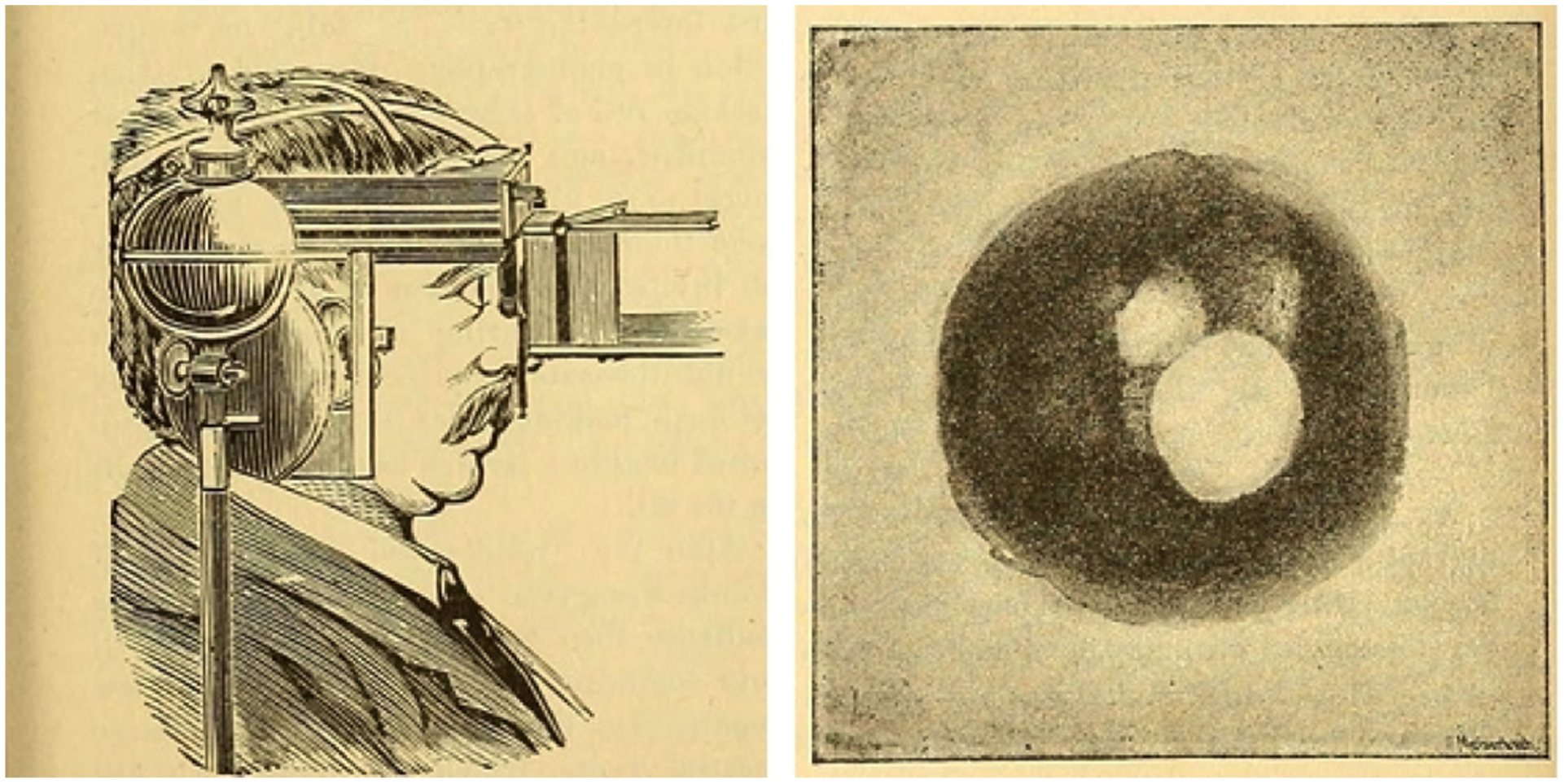
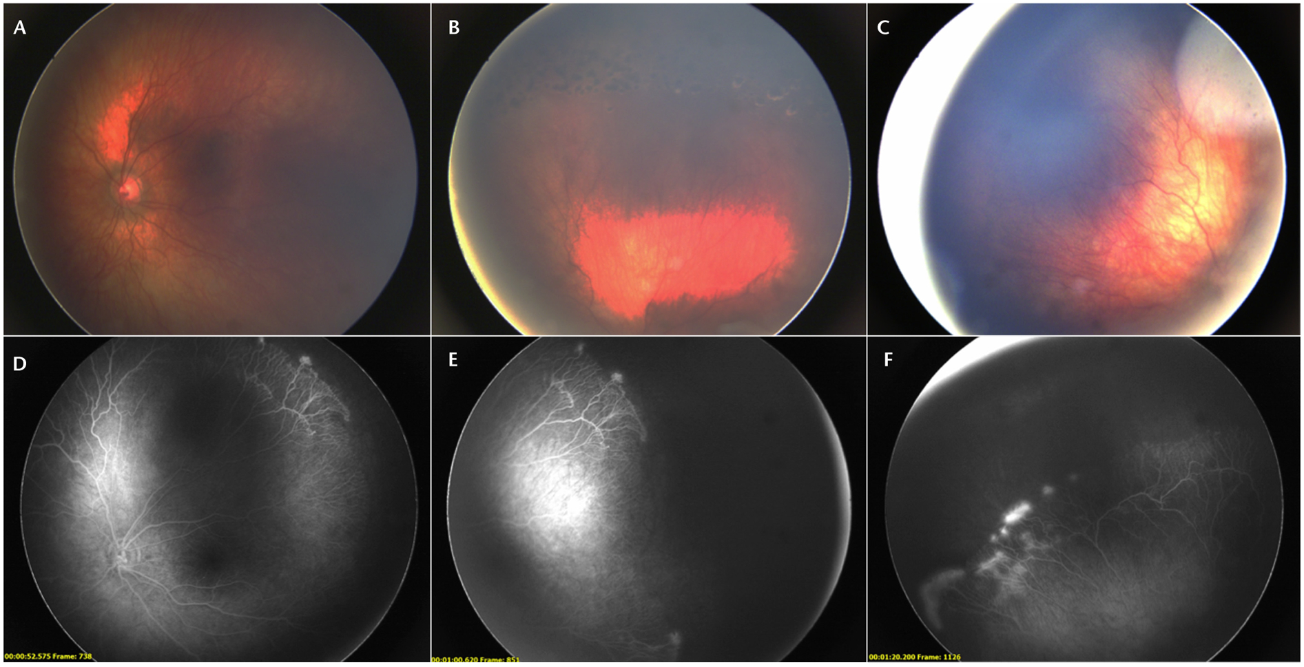
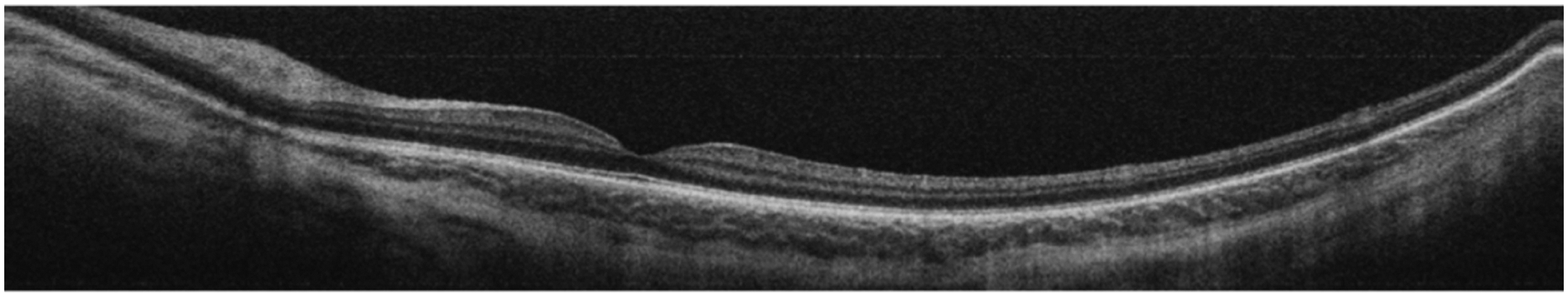
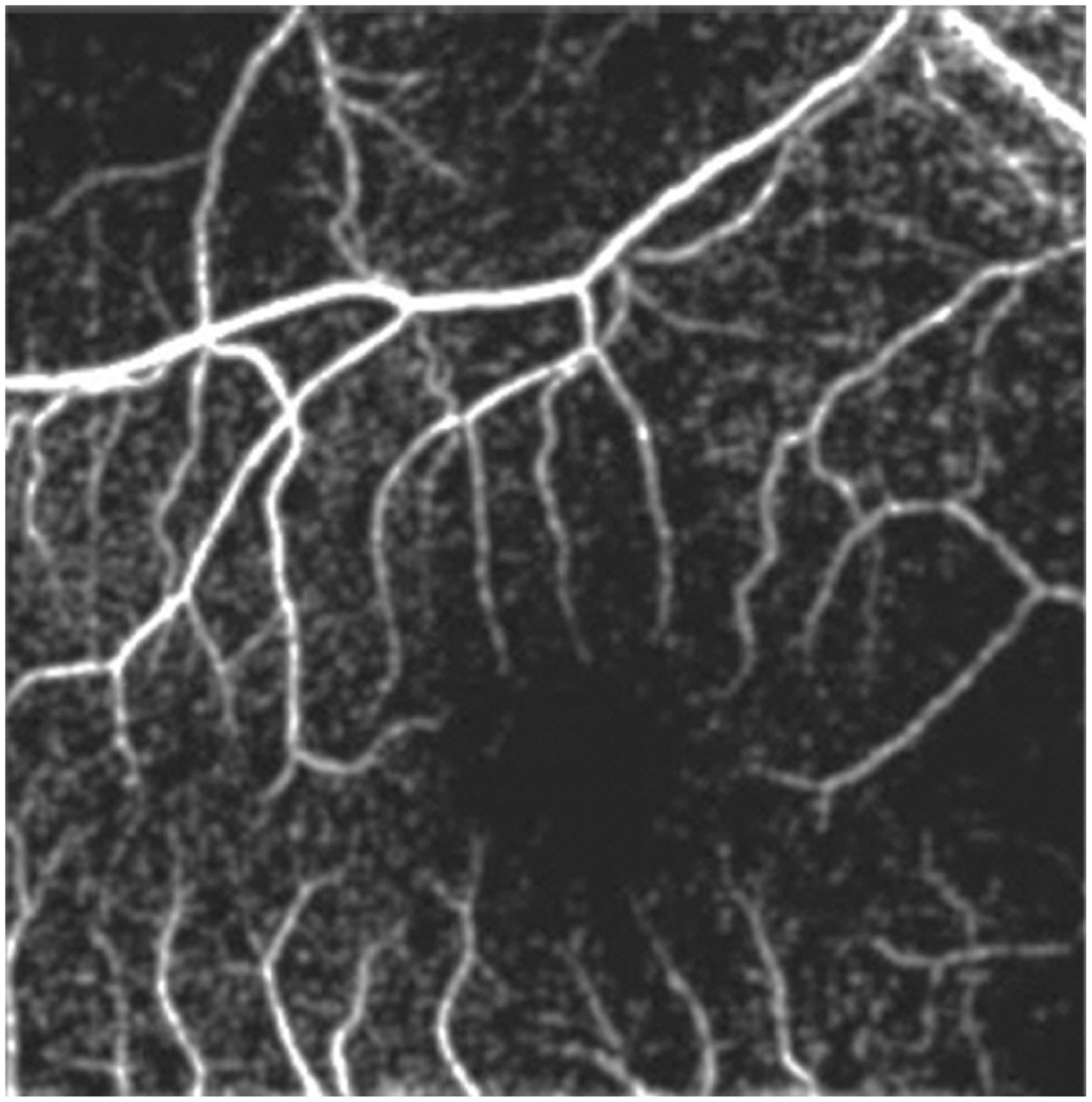
Retinopathy of prematurity (ROP) is a leading cause of preventable childhood blindness worldwide. Barriers to ROP screening and difficulties with subsequent evaluation and management include poor access to care, lack of physicians trained in ROP, and issues with objective documentation. Digital retinal imaging can help address these barriers and improve our knowledge of the pathophysiology of the disease. Advancements in technology have led to new, non-mydriatic and mydriatic cameras with wider fields of view as well as devices that can simultaneously incorporate fluorescein angiography, optical coherence tomography (OCT), and OCT angiography. Image analysis in ROP is also being employed through smartphones and computer-based software. Telemedicine programs in the United States and worldwide have utilized imaging to extend ROP screening to infants in remote areas and have shown that digital retinal imaging can be reliable, accurate, and cost-effective. In addition, tele-education programs are also using digital retinal images to increase the number of healthcare providers trained in ROP. Although indirect ophthalmoscopy is still an important skill for screening, digital retinal imaging holds promise for more widespread screening and management of ROP.
Keywords: imaging; retinopathy of prematurity; telemedicine.
Copyright 2019 Asia-Pacific Academy of Ophthalmology.
Figures
References
-
- International Committee for the Classification of Retinopathy of Prematurity. The International Classification of Retinopathy of Prematurity revisited. Arch Ophthalmol. 2005;123:991–999. - PubMed
-
- Early Treatment For Retinopathy Of Prematurity Cooperative Group. Revised indications for the treatment of retinopathy of prematurity: results of the early treatment for retinopathy of prematurity randomized trial. Arch Ophthalmol. 2003;121:1684–1694. - PubMed
-
- Multicenter trial of cryotherapy for retinopathy of prematurity: preliminary results. Cryotherapy for Retinopathy of Prematurity Cooperative Group. Pediatrics. 1988;81:697–706. - PubMed
-
- Vural A, Perente İ, Onur İU, et al. Efficacy of intravitreal aflibercept monotherapy in retinopathy of prematurity evaluated by periodic fluorescence angiography and optical coherence tomography. Int Ophthalmol. 2018. November 26 Epub ahead of print. - PubMed
