. 2019 Jul;292(1):226-234.
doi: 10.1148/radiol.2019182135. Epub 2019 Apr 30.

LI-RADS Treatment Response Algorithm: Performance and Diagnostic Accuracy

Affiliations

LI-RADS Treatment Response Algorithm: Performance and Diagnostic Accuracy

Erin L Shropshire et al. Radiology. 2019 Jul.

Abstract

Background In 2017, the Liver Imaging Reporting and Data System (LI-RADS) included an algorithm for the assessment of hepatocellular carcinoma (HCC) treated with local-regional therapy. The aim of the algorithm was to enable standardized evaluation of treatment response to guide subsequent therapy. However, the performance of the algorithm has not yet been validated in the literature. Purpose To evaluate the performance of the LI-RADS 2017 Treatment Response algorithm for assessing the histopathologic viability of HCC treated with bland arterial embolization. Materials and Methods This retrospective study included patients who underwent bland arterial embolization for HCC between 2006 and 2016 and subsequent liver transplantation. Three radiologists independently assessed all treated lesions by using the CT/MRI LI-RADS 2017 Treatment Response algorithm. Radiology and posttransplant histopathology reports were then compared. Lesions were categorized on the basis of explant pathologic findings as either completely (100%) or incompletely (<100%) necrotic, and performance characteristics and predictive values for the LI-RADS Treatment Response (LR-TR) Viable and Nonviable categories were calculated for each reader. Interreader association was calculated by using the Fleiss κ. Results A total of 45 adults (mean age, 57.1 years ± 8.2; 13 women) with 63 total lesions were included. For predicting incomplete histopathologic tumor necrosis, the accuracy of the LR-TR Viable category for the three readers was 60%-65%, and the positive predictive value was 86%-96%. For predicting complete histopathologic tumor necrosis, the accuracy of the LR-TR Nonviable category was 67%-71%, and the negative predictive value was 81%-87%. By consensus, 17 (27%) of 63 lesions were categorized as LR-TR Equivocal, and 12 of these lesions were incompletely necrotic. Interreader association for the LR-TR category was moderate (κ = 0.55; 95% confidence interval: 0.47, 0.67). Conclusion The Liver Imaging Reporting and Data System 2017 Treatment Response algorithm had high predictive value and moderate interreader association for the histopathologic viability of hepatocellular carcinoma treated with bland arterial embolization when lesions were assessed as Viable or Nonviable. © RSNA, 2019 Online supplemental material is available for this article. See also the editorial by Gervais in this issue.


Figures
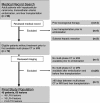
Figure 1:
Figure 1:
Study flowchart shows a summary of the inclusion and exclusion criteria and final study population.
Figure 2a:
Figure 2a:
Images in 65-year-old man with cirrhosis due to nonalcoholic steatohepatitis. (a, b) Images obtained at 3.0-T MRI performed by using a three-dimensional (3D) T1-weighted gradient-recalled echo sequence (repetition time msec/echo time msec, 4.2/1.2; flip angle, 15°; matrix, 320 × 248). Axial (a) arterial and (b) portal venous phase images obtained before embolization show a 42-mm hyperenhancing lesion (arrow) with washout that was deemed a Liver Imaging Reporting and Data System, or LI-RADS, category 5 lesion. (c, d) Images obtained at 1.5-T MRI performed by using a 3D T1-weighted gradient-recalled echo sequence (4.1/2.0; flip angle, 10°; matrix, 256 × 205). On pretransplant axial (c) arterial and (d) portal venous phase images obtained 71 days after embolization, the lesion (arrow) had increased in size to 51 mm. It demonstrated mass-like irregular arterial phase hyperenhancement. This lesion was categorized as LR-TR Viable by all three readers. At histologic examination, this lesion was less than 50% necrotic.
Figure 2b:
Figure 2b:
Images in 65-year-old man with cirrhosis due to nonalcoholic steatohepatitis. (a, b) Images obtained at 3.0-T MRI performed by using a three-dimensional (3D) T1-weighted gradient-recalled echo sequence (repetition time msec/echo time msec, 4.2/1.2; flip angle, 15°; matrix, 320 × 248). Axial (a) arterial and (b) portal venous phase images obtained before embolization show a 42-mm hyperenhancing lesion (arrow) with washout that was deemed a Liver Imaging Reporting and Data System, or LI-RADS, category 5 lesion. (c, d) Images obtained at 1.5-T MRI performed by using a 3D T1-weighted gradient-recalled echo sequence (4.1/2.0; flip angle, 10°; matrix, 256 × 205). On pretransplant axial (c) arterial and (d) portal venous phase images obtained 71 days after embolization, the lesion (arrow) had increased in size to 51 mm. It demonstrated mass-like irregular arterial phase hyperenhancement. This lesion was categorized as LR-TR Viable by all three readers. At histologic examination, this lesion was less than 50% necrotic.
Figure 2c:
Figure 2c:
Images in 65-year-old man with cirrhosis due to nonalcoholic steatohepatitis. (a, b) Images obtained at 3.0-T MRI performed by using a three-dimensional (3D) T1-weighted gradient-recalled echo sequence (repetition time msec/echo time msec, 4.2/1.2; flip angle, 15°; matrix, 320 × 248). Axial (a) arterial and (b) portal venous phase images obtained before embolization show a 42-mm hyperenhancing lesion (arrow) with washout that was deemed a Liver Imaging Reporting and Data System, or LI-RADS, category 5 lesion. (c, d) Images obtained at 1.5-T MRI performed by using a 3D T1-weighted gradient-recalled echo sequence (4.1/2.0; flip angle, 10°; matrix, 256 × 205). On pretransplant axial (c) arterial and (d) portal venous phase images obtained 71 days after embolization, the lesion (arrow) had increased in size to 51 mm. It demonstrated mass-like irregular arterial phase hyperenhancement. This lesion was categorized as LR-TR Viable by all three readers. At histologic examination, this lesion was less than 50% necrotic.
Figure 2d:
Figure 2d:
Images in 65-year-old man with cirrhosis due to nonalcoholic steatohepatitis. (a, b) Images obtained at 3.0-T MRI performed by using a three-dimensional (3D) T1-weighted gradient-recalled echo sequence (repetition time msec/echo time msec, 4.2/1.2; flip angle, 15°; matrix, 320 × 248). Axial (a) arterial and (b) portal venous phase images obtained before embolization show a 42-mm hyperenhancing lesion (arrow) with washout that was deemed a Liver Imaging Reporting and Data System, or LI-RADS, category 5 lesion. (c, d) Images obtained at 1.5-T MRI performed by using a 3D T1-weighted gradient-recalled echo sequence (4.1/2.0; flip angle, 10°; matrix, 256 × 205). On pretransplant axial (c) arterial and (d) portal venous phase images obtained 71 days after embolization, the lesion (arrow) had increased in size to 51 mm. It demonstrated mass-like irregular arterial phase hyperenhancement. This lesion was categorized as LR-TR Viable by all three readers. At histologic examination, this lesion was less than 50% necrotic.
Figure 3a:
Figure 3a:
Images in 68-year-old man with cirrhosis secondary to chronic hepatitis C infection. (a, b) Images obtained at 3.0-T MRI performed by using a three-dimensional (3D) T1-weighted gradient-recalled echo sequence (repetition time msec/echo time msec, 4.2/1.3; flip angle, 15°; matrix, 320 × 256). Axial (a) arterial and (b) portal venous phase images obtained before embolization show a 13-mm hyperenhancing lesion (arrow) with washout and a capsule that was deemed a Liver Imaging Reporting and Data System, or LI-RADS, category 5 lesion. (c, d) Images obtained at 1.5-T MRI performed by using a 3D T1-weighted gradient-recalled echo sequence (4.4/2.1; flip angle, 12°; matrix, 256 × 192). On pretransplant axial (c) arterial and (d) portal venous phase images obtained 227 days after embolization, there was no residual arterial phase hyperenhancement or washout associated with the lesion (arrow). This lesion was categorized as LR-TR Nonviable by all three readers. At histologic examination, this lesion was 100% necrotic.
Figure 3b:
Figure 3b:
Images in 68-year-old man with cirrhosis secondary to chronic hepatitis C infection. (a, b) Images obtained at 3.0-T MRI performed by using a three-dimensional (3D) T1-weighted gradient-recalled echo sequence (repetition time msec/echo time msec, 4.2/1.3; flip angle, 15°; matrix, 320 × 256). Axial (a) arterial and (b) portal venous phase images obtained before embolization show a 13-mm hyperenhancing lesion (arrow) with washout and a capsule that was deemed a Liver Imaging Reporting and Data System, or LI-RADS, category 5 lesion. (c, d) Images obtained at 1.5-T MRI performed by using a 3D T1-weighted gradient-recalled echo sequence (4.4/2.1; flip angle, 12°; matrix, 256 × 192). On pretransplant axial (c) arterial and (d) portal venous phase images obtained 227 days after embolization, there was no residual arterial phase hyperenhancement or washout associated with the lesion (arrow). This lesion was categorized as LR-TR Nonviable by all three readers. At histologic examination, this lesion was 100% necrotic.
Figure 3c:
Figure 3c:
Images in 68-year-old man with cirrhosis secondary to chronic hepatitis C infection. (a, b) Images obtained at 3.0-T MRI performed by using a three-dimensional (3D) T1-weighted gradient-recalled echo sequence (repetition time msec/echo time msec, 4.2/1.3; flip angle, 15°; matrix, 320 × 256). Axial (a) arterial and (b) portal venous phase images obtained before embolization show a 13-mm hyperenhancing lesion (arrow) with washout and a capsule that was deemed a Liver Imaging Reporting and Data System, or LI-RADS, category 5 lesion. (c, d) Images obtained at 1.5-T MRI performed by using a 3D T1-weighted gradient-recalled echo sequence (4.4/2.1; flip angle, 12°; matrix, 256 × 192). On pretransplant axial (c) arterial and (d) portal venous phase images obtained 227 days after embolization, there was no residual arterial phase hyperenhancement or washout associated with the lesion (arrow). This lesion was categorized as LR-TR Nonviable by all three readers. At histologic examination, this lesion was 100% necrotic.
Figure 3d:
Figure 3d:
Images in 68-year-old man with cirrhosis secondary to chronic hepatitis C infection. (a, b) Images obtained at 3.0-T MRI performed by using a three-dimensional (3D) T1-weighted gradient-recalled echo sequence (repetition time msec/echo time msec, 4.2/1.3; flip angle, 15°; matrix, 320 × 256). Axial (a) arterial and (b) portal venous phase images obtained before embolization show a 13-mm hyperenhancing lesion (arrow) with washout and a capsule that was deemed a Liver Imaging Reporting and Data System, or LI-RADS, category 5 lesion. (c, d) Images obtained at 1.5-T MRI performed by using a 3D T1-weighted gradient-recalled echo sequence (4.4/2.1; flip angle, 12°; matrix, 256 × 192). On pretransplant axial (c) arterial and (d) portal venous phase images obtained 227 days after embolization, there was no residual arterial phase hyperenhancement or washout associated with the lesion (arrow). This lesion was categorized as LR-TR Nonviable by all three readers. At histologic examination, this lesion was 100% necrotic.
Figure 4a:
Figure 4a:
Images in 60-year-old woman with cirrhosis due to chronic hepatitis C infection. (a, b) Images obtained at 1.5-T MRI performed by using a three-dimensional (3D) T1-weighted gradient-recalled echo sequence (repetition time msec/echo time msec, 7.5/2.4; flip angle, 10°; matrix, 256 × 170). Axial (b) arterial and (b) portal venous phase images obtained before embolization show a 21-mm hyperenhancing lesion (arrow) with washout that was deemed a Liver Imaging Reporting and Data System, or LI-RADS, category 5 lesion. (c, d) Images obtained at 1.5-T MRI performed by using a 3D T1-weighted gradient-recalled echo sequence (7.5/2.4; flip angle, 10°; matrix, 256 × 170). On pretransplant axial (c) arterial and (d) portal venous phase images obtained 168 days after embolization, the lesion (arrow) had decreased in size to 7 mm. Two of three readers assessed indeterminate arterial phase hyperenhancement, while one reader assessed focal mass-like arterial hyperenhancement. This lesion was categorized as LR-TR Equivocal by two readers and as LR-TR Viable by one reader. At histologic examination, this lesion was less than 50% necrotic.
Figure 4b:
Figure 4b:
Images in 60-year-old woman with cirrhosis due to chronic hepatitis C infection. (a, b) Images obtained at 1.5-T MRI performed by using a three-dimensional (3D) T1-weighted gradient-recalled echo sequence (repetition time msec/echo time msec, 7.5/2.4; flip angle, 10°; matrix, 256 × 170). Axial (b) arterial and (b) portal venous phase images obtained before embolization show a 21-mm hyperenhancing lesion (arrow) with washout that was deemed a Liver Imaging Reporting and Data System, or LI-RADS, category 5 lesion. (c, d) Images obtained at 1.5-T MRI performed by using a 3D T1-weighted gradient-recalled echo sequence (7.5/2.4; flip angle, 10°; matrix, 256 × 170). On pretransplant axial (c) arterial and (d) portal venous phase images obtained 168 days after embolization, the lesion (arrow) had decreased in size to 7 mm. Two of three readers assessed indeterminate arterial phase hyperenhancement, while one reader assessed focal mass-like arterial hyperenhancement. This lesion was categorized as LR-TR Equivocal by two readers and as LR-TR Viable by one reader. At histologic examination, this lesion was less than 50% necrotic.
Figure 4c:
Figure 4c:
Images in 60-year-old woman with cirrhosis due to chronic hepatitis C infection. (a, b) Images obtained at 1.5-T MRI performed by using a three-dimensional (3D) T1-weighted gradient-recalled echo sequence (repetition time msec/echo time msec, 7.5/2.4; flip angle, 10°; matrix, 256 × 170). Axial (b) arterial and (b) portal venous phase images obtained before embolization show a 21-mm hyperenhancing lesion (arrow) with washout that was deemed a Liver Imaging Reporting and Data System, or LI-RADS, category 5 lesion. (c, d) Images obtained at 1.5-T MRI performed by using a 3D T1-weighted gradient-recalled echo sequence (7.5/2.4; flip angle, 10°; matrix, 256 × 170). On pretransplant axial (c) arterial and (d) portal venous phase images obtained 168 days after embolization, the lesion (arrow) had decreased in size to 7 mm. Two of three readers assessed indeterminate arterial phase hyperenhancement, while one reader assessed focal mass-like arterial hyperenhancement. This lesion was categorized as LR-TR Equivocal by two readers and as LR-TR Viable by one reader. At histologic examination, this lesion was less than 50% necrotic.
Figure 4d:
Figure 4d:
Images in 60-year-old woman with cirrhosis due to chronic hepatitis C infection. (a, b) Images obtained at 1.5-T MRI performed by using a three-dimensional (3D) T1-weighted gradient-recalled echo sequence (repetition time msec/echo time msec, 7.5/2.4; flip angle, 10°; matrix, 256 × 170). Axial (b) arterial and (b) portal venous phase images obtained before embolization show a 21-mm hyperenhancing lesion (arrow) with washout that was deemed a Liver Imaging Reporting and Data System, or LI-RADS, category 5 lesion. (c, d) Images obtained at 1.5-T MRI performed by using a 3D T1-weighted gradient-recalled echo sequence (7.5/2.4; flip angle, 10°; matrix, 256 × 170). On pretransplant axial (c) arterial and (d) portal venous phase images obtained 168 days after embolization, the lesion (arrow) had decreased in size to 7 mm. Two of three readers assessed indeterminate arterial phase hyperenhancement, while one reader assessed focal mass-like arterial hyperenhancement. This lesion was categorized as LR-TR Equivocal by two readers and as LR-TR Viable by one reader. At histologic examination, this lesion was less than 50% necrotic.

Comment in

References

    1. Fraum TJ, Tsai R, Rohe E, et al. Differentiation of hepatocellular carcinoma from other hepatic malignancies in patients at risk: diagnostic performance of the liver imaging reporting and data system version 2014 . Radiology 2018. ; 286 ( 1 ): 158 – 172 . - PubMed
    1. Ferlay J, Soerjomataram I, Dikshit R, et al. Cancer incidence and mortality worldwide: sources, methods and major patterns in GLOBOCAN 2012 . Int J Cancer 2015. ; 136 ( 5 ): E359 – E386 . - PubMed
    1. Kulik L, Heimbach JK, Zaiem F, et al. Therapies for patients with hepatocellular carcinoma awaiting liver transplantation: a systematic review and meta-analysis . Hepatology 2018. ; 67 ( 1 ): 381 – 400 . - PubMed
    1. Dhanasekaran R, Khanna V, Kooby DA, et al. The effectiveness of locoregional therapies versus supportive care in maintaining survival within the Milan criteria in patients with hepatocellular carcinoma . J Vasc Interv Radiol 2010. ; 21 ( 8 ): 1197 – 1204 ; quiz 204 . - PubMed
    1. Gaba RC, Lokken RP, Hickey RM, et al. Quality improvement guidelines for transarterial chemoembolization and embolization of hepatic malignancy . J Vasc Interv Radiol 2017. ; 28 ( 9 ): 1210 – 1223 . e3 . - PubMed

Publication types

MeSH terms