. 2019 Apr 30;129(7):2878-2887.
doi: 10.1172/JCI127195.

Aberrant splicing contributes to severe α-spectrin-linked congenital hemolytic anemia

Affiliations

Aberrant splicing contributes to severe α-spectrin-linked congenital hemolytic anemia

Patrick G Gallagher et al. J Clin Invest. .

Abstract

The etiology of severe hemolytic anemia in most patients with recessive hereditary spherocytosis (rHS) and the related disorder hereditary pyropoikilocytosis (HPP) is unknown. Whole exome sequencing of DNA from probands of 24 rHS or HPP kindreds identified numerous mutations in erythrocyte membrane α-spectrin (SPTA1). Twenty-eight mutations were novel, with null alleles frequently found in trans to missense mutations. No mutations were identified in a third of SPTA1 alleles (17/48). Whole genome sequencing revealed linkage disequilibrium between the common rHS-linked α-spectrinBug Hill polymorphism and a rare intron 30 variant in all 17 mutation-negative alleles. In vitro minigene studies and in vivo splicing analyses revealed the intron 30 variant changes a weak alternate branch point (BP) to a strong BP. This change leads to increased utilization of an alternate 3' splice acceptor site, perturbing normal α-spectrin mRNA splicing and creating an elongated mRNA transcript. In vivo mRNA stability studies revealed the newly created termination codon in the elongated transcript activates nonsense mediated decay leading to spectrin deficiency. These results demonstrate a unique mechanism of human genetic disease contributes to the etiology of a third of cases of rHS, facilitating diagnosis and treatment of severe anemia, and identifying a new target for therapeutic manipulation.

Keywords: Genetic diseases; Genetics; Hematology.


Conflict of interest statement

Conflict of interest: The authors have declared that no conflict of interest exists.

Figures

Figure 1
Figure 1. Haplotyping at the SPTA1 locus.
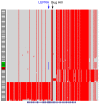
Haplotype analysis around the SPTA1 locus of all 71 alleles with the αBH variant, Ala970Asp, in the 1000 Genomes 2015 database and 2 rHS patients heterozygous for the αBH variant. The αBH allele is denoted in black and the rare intron 30 variant, the αLEPRA allele, is denoted in blue. The HG19 reference sequence is denoted in gray and the nonreference sequence is shown in red. The SPTA1 gene is shown at the bottom. The 4 individuals in the 2015 1000 Genomes database and both rHS patients who carry the αLEPRA allele are shown in green and red, respectively.
Figure 2
Figure 2. An elongated transcript of intron 30 of the SPTA1 gene.
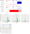
(A) Normalized RNA-seq profiles of the SPTA1 intron 30 region in human WT, primary erythroid cells, and K562 cells are shown. Estimated percentages of elongated, partial intron 30–containing mRNA transcripts based on splice junction reads are shown. (B) A Sashimi plot demonstrates utilization of splice sites in intron-spanning reads.
Figure 3
Figure 3. Computational analyses of intron 30 splicing of the human SPTA1 gene.
A schematic of cis sequences and BP usage in WT (A) and αLEPRA SPTA1 (B) alleles. In WT cells, BP1 is primarily utilized, producing large amounts of correctly spliced, in-frame mRNA transcripts (blue), whereas utilization of a weak, alternate BP2 produces a small amount of elongated transcript that leads to a frameshift and a novel termination codon (red). The αLEPRA mutation, which changes BP2 from a weak alternate BP to a strong alternate BP, leads to increased utilization of BP2, producing large amounts of elongated transcript with decreased utilization of BP1. This leads to production of a small amount of correctly spliced transcript. (C) Branchpointer, an algorithm that predicts branch points, shows both BP1 and BP2-WT have low BP probability scores (0.51 and 0.67, respectively), while the αLEPRA variant markedly improves the probability score of BP2-L (0.94). (D) These observations are true when comparing intron 30 BP probability scores to the predicted BP probability scores of the BPs of 8180 introns of 1000 highly expressed erythroid genes.
Figure 4
Figure 4. Minigene studies of α-spectrin intron 30 and the αLEPRA variant.
(A) Partial sequence of intron 30 of the SPTA1 gene, showing the location of the αLEPRA variant. (B) Each minigene construct used in minigene assays includes the ANK1 erythroid promoter, a fragment of SPTA1 genomic DNA inserted into intron 2 of the HBG1 gene, and the HBG1 3′ untranslated region and polyA signal. The hybrid HBG1-SPTA1 transcripts derived from minigenes are shown, either WT or elongated, with the locations of Taqman probes utilized to detect total spectrin (T bar) or the unique insert of the elongated transcript (E bar). (C) Minigene results. The specific sequences utilized in minigene constructs are shown. Percentages of elongated α-spectrin transcript over total α-spectrin transcript are shown in the second column from right. The adjusted P value of the difference from WT is shown on the right.
Figure 5
Figure 5. Analyses of nonsense-mediated decay.
The influence of NMD on the stability of the elongated α-spectrin transcript in WT K562 cells and in K562 cells rendered homozygous for the αLEPRA allele. (A) After treatment with the NMD inhibitor emetine (E) or cycloheximide (C), or both, the amounts of total α-spectrin and the elongated α-spectrin transcript were determined by real-time RT-PCR with fluorescent Taqman probes. After NMD inhibition, amounts of elongated α-spectrin transcript were increased in αLEPRA cells (adjusted P values 0.0014, 0.0027, and 0.0014 for WT vs. cycloheximide, emetine, and cycloheximide + emetine, respectively) and to a lesser extent in WT cells (adjusted P values 0.084, 0.012, and 0.0047, respectively). (B) Minigene assay of NMD. Two nucleotides were inserted into the polypyrimidine tract of the primary 3′ acceptor site of exon 30 in the minigene model to place the elongated transcript in-frame. As predicted, NMD was abrogated and the amounts of elongated α-spectrin transcript from both the WT and the αLEPRA minigene constructs significantly increased. Three biologic samples each were tested in 3 independent experiments with the mean calculated from 9 values. Statistical significance was determined by 2-tailed Student’s t test.

Comment in

  • Anemia lurking in introns.
    Mohandas N. Mohandas N. J Clin Invest. 2019 Jun 4;129(7):2655-2657. doi: 10.1172/JCI129443. eCollection 2019 Jun 4. J Clin Invest. 2019. PMID: 31162137 Free PMC article.

References

    1. Agre P, Orringer EP, Bennett V. Deficient red-cell spectrin in severe, recessively inherited spherocytosis. N Engl J Med. 1982;306(19):1155–1161. doi: 10.1056/NEJM198205133061906. - DOI - PubMed
    1. Agre P, Casella JF, Zinkham WH, McMillan C, Bennett V. Partial deficiency of erythrocyte spectrin in hereditary spherocytosis. Nature. 1985;314(6009):380–383. doi: 10.1038/314380a0. - DOI - PubMed
    1. Tse WT, et al. Amino-acid substitution in alpha-spectrin commonly coinherited with nondominant hereditary spherocytosis. Am J Hematol. 1997;54(3):233–241. doi: 10.1002/(SICI)1096-8652(199703)54:3<233::AID-AJH10>3.0.CO;2-E. - DOI - PubMed
    1. Mohandas N. Inherited hemolytic anemia: a possessive beginner’s guide. Hematology Am Soc Hematol Educ Program. 2018;2018(1):377–381. - PMC - PubMed
    1. Gallagher PG. Abnormalities of the erythrocyte membrane. Pediatr Clin North Am. 2013;60(6):1349–1362. doi: 10.1016/j.pcl.2013.09.001. - DOI - PMC - PubMed

Publication types