. 2019 Apr 30;5(11):e124430.
doi: 10.1172/jci.insight.124430.

Sensitive and adaptable pharmacological control of CAR T cells through extracellular receptor dimerization

Affiliations

Sensitive and adaptable pharmacological control of CAR T cells through extracellular receptor dimerization

Wai-Hang Leung et al. JCI Insight. .

Abstract

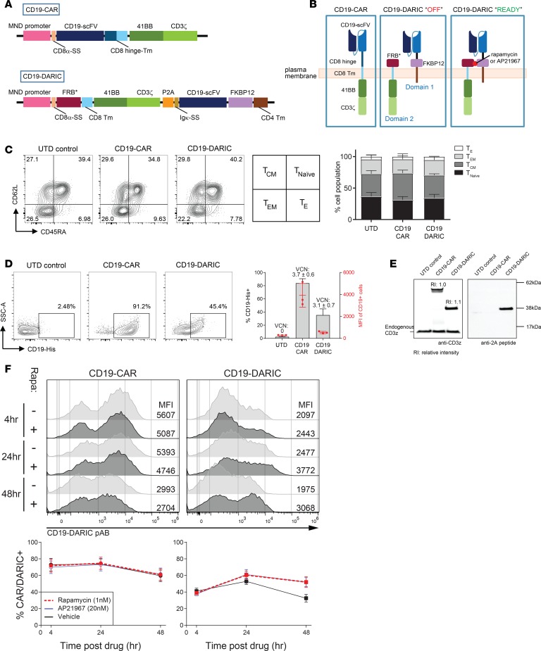
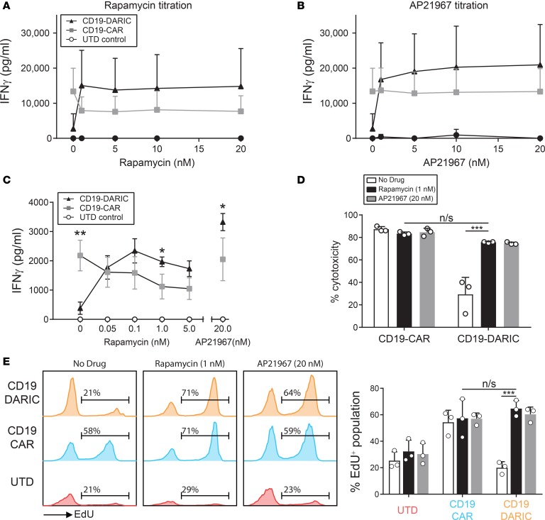
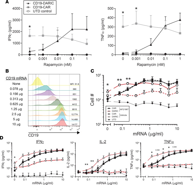
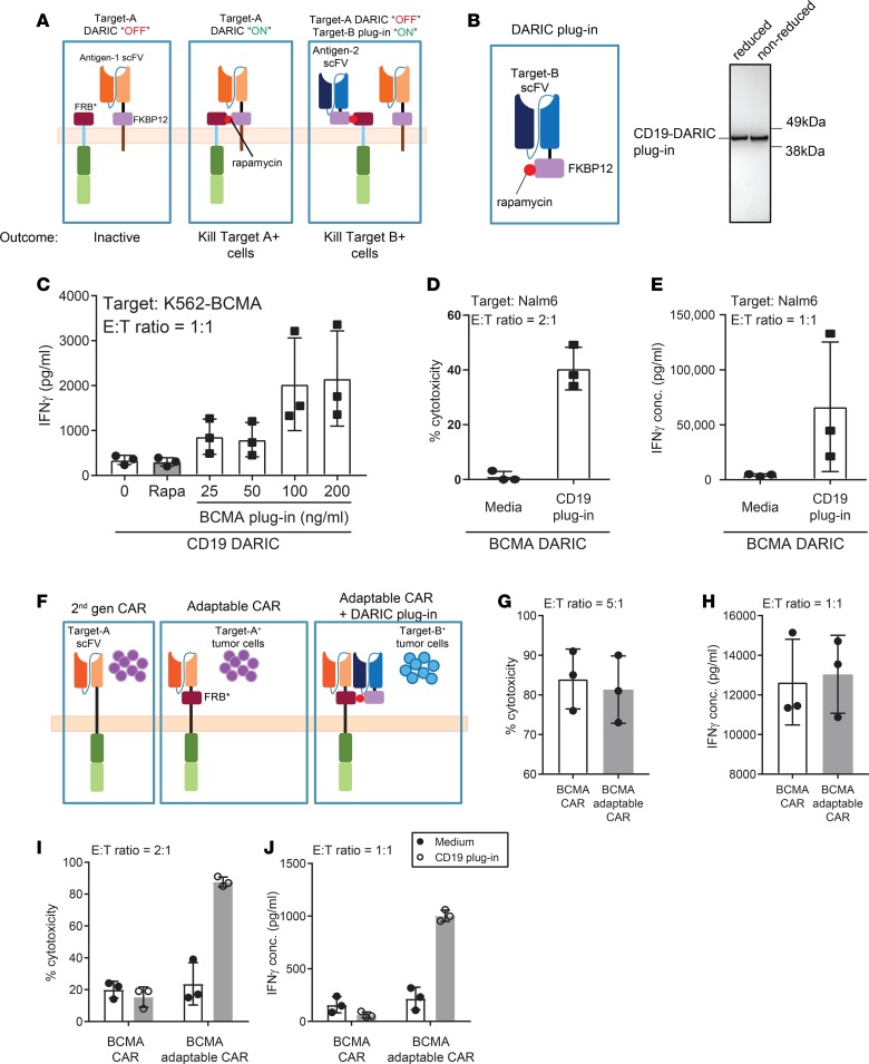
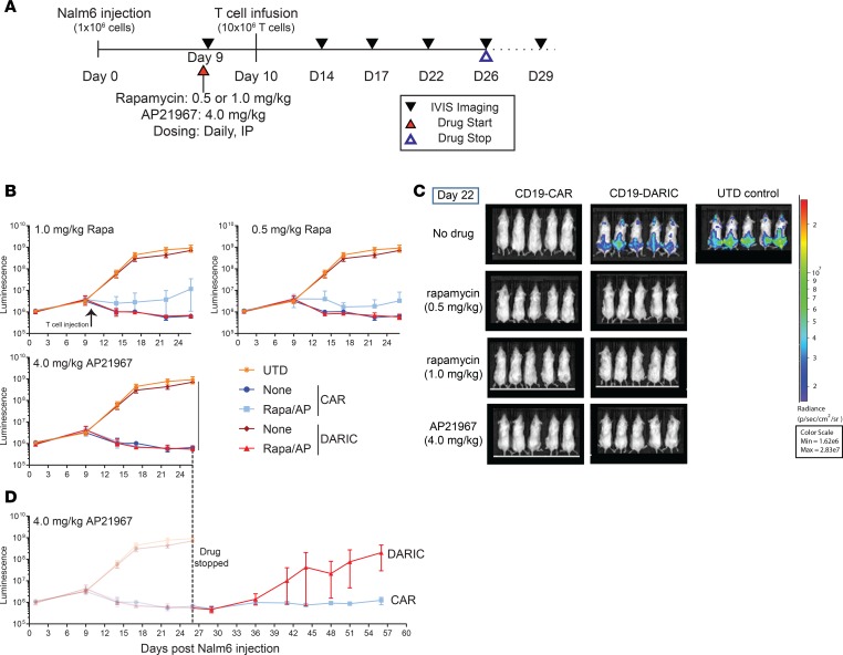
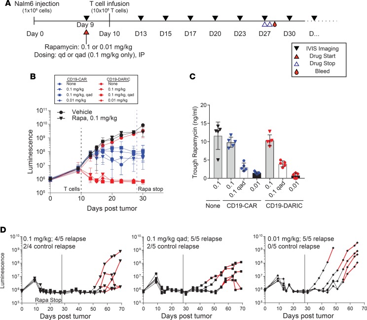
Chimeric antigen receptor (CAR) T cell therapies have achieved promising outcomes in several cancers, however more challenging oncology indications may necessitate advanced antigen receptor designs and functions. Here we describe a bipartite receptor system comprised of separate antigen targeting and signal transduction polypeptides, each containing an extracellular dimerization domain. We demonstrate that T cell activation remains antigen dependent but can only be achieved in the presence of a dimerizing drug, rapamycin. Studies performed in vitro and in xenograft mouse models illustrate equivalent to superior anti-tumor potency compared to currently used CAR designs, and at rapamycin concentrations well below immunosuppressive levels. We further show that the extracellular positioning of the dimerization domains enables the administration of recombinant re-targeting modules, potentially extending antigen targeting. Overall, this novel regulatable CAR design has exquisite drug sensitivity, provides robust anti-tumor responses, and is uniquely flexible for multiplex antigen targeting or retargeting, which may further assist the development of safe, potent and durable T cell therapeutics.

Keywords: Cancer immunotherapy; Gene therapy; Leukemias; Oncology; Therapeutics.


Conflict of interest statement

Conflict of interest: All authors, except BRB, are employees and shareholders of bluebird bio. WHL, JG, MTC, JJ, and AA are co-inventors on patent applications describing the DARIC technology. BRB receives remuneration as an advisor to Kamon Pharmaceuticals, Inc, Five Prime Therapeutics Inc, Regeneron Pharmaceuticals, Magenta Therapeutics and BlueRock Therapeuetics; research support from Fate Therapeutics, RXi Pharmaceuticals, Alpine Immune Sciences, Inc, Abbvie Inc., Leukemia and Lymphoma Society, Childrens’ Cancer Research Fund, KidsFirst Fund and is a co-founder of Tmunity.

Figures

Figure 1
Figure 1. CD19-DARIC transduction results in normal T cell development.
(A) Schematic of the lentiviral vectors used in the study. SS, signal sequence; TM, transmembrane domain. (B) The CD19-DARIC T cells are inactive in the absence of drug (“OFF”) and addition of dimerizing agent brings the 2 domains together to turn the receptor “ON”. (C) T cells were transduced, expanded, and phenotyped after 10 days of expansion with CD62L and CD45RA. The T cell memory and naive compartments were identified on the basis of relative marker expression and the summary data are shown on the right. (D) The transduced cells were stained with fluorescently conjugated CD19-His antigen and analyzed by FACS. Summary of 3 donors is shown on the right, with the MFI values in red and the VCN values for each sample annotated above. (E) Western blot analysis of T cell lysates using CD3ζ- and 2A-specific antibodies. Relative intensity was determined using LICOR Western blot analysis software. (F) CD19-CAR or CD19-DARIC T cells were cultured for the indicated amount of time in the presence or absence of rapamycin. The expression of the CAR/DARIC construct was analyzed by staining with rabbit polyclonal anti–CD19 complex antibody. The summary of the staining data is shown below. Data are representative of at least 3 separate experiments, with 3 unique donors per experiment.
Figure 2
Figure 2. CD19-DARIC T cells are tumor reactive solely in the presence of a dimerization drug.
(AC) The CD19-CAR and CD19-DARIC T cells were cultured at a 1:1 ratio with fluorescent Nalm-6 target cells with or without different concentrations of either rapamycin or AP21967. Supernatant was collected 24 hours after culture initiation and cytokine levels were analyzed using IFN-γ–specific ELISA (A and B) or iQue QBead assay (C) (n = 3). *P < 0.05; **P < 0.01 as determined by a 2-tailed unpaired Student’s t test. (D) The percentage cytotoxicity was determined by analyzing the ratio of fluorescent Nalm-6 cells to antigen-naive K562 cells following a 24-hour coculture with CAR or DARIC T cells. (E) The T cells were cocultured with Nalm-6 for 72 hours in the indicated conditions. Modified EdU was added and the cells were cultured for another 24 hours prior to analysis of EdU incorporation. The percentage of EdU+ cells represents the proportion of cells that underwent DNA synthesis in the prior 24 hours. ***P < 0.001 using 1-way ANOVA with Dunnett’s test for multiparameter comparison to CD19-DARIC T cells cultured with rapamycin. n/s, not significant.
Figure 3
Figure 3. CD19-DARIC T cells are potent even at low rapamycin concentrations and low antigen expression.
(A) CD19-DARIC or CD19-CAR T cells were cultured with Nalm-6 cells for 24 hours in the presence of different concentrations of rapamycin. Cytokine production was analyzed using iQue QBeads. Data points represent 3 donors. (B) K562 cells were transfected with in vitro–transcribed mRNA encoding the CD19 antigen. The transfected cells were cultured for 24 hours and CD19 expression was analyzed by flow cytometry. The amount of CD19 mRNA for each transfection is shown on the left, and the CD19 MFI value is listed on the right. (C) CD19-transfected K562 cells were cultured with CD19-DARIC (red) or CD19-CAR (black) T cells in the presence of 20 nM AP21967. After a 24-hour incubation period, supernatant was collected for cytokine analysis and cells were cultured for an additional 72 hours (4 days total coculture). At the conclusion of the coculture period, the number of T cells in each well was counted. The dashed lines represent samples cultured without AP21967, while solid lines represent samples cultured in the presence of 20 nM AP21967. The gray line indicates untransduced control. (D) Cytokine production in 24-hour supernatants from C was analyzed using iQue QBeads. Data points represent 3 unique donors. *P < 0.05; **P < 0.01 as determined by a 2-tailed, unpaired Student’s t test comparing AP21967-treated CD19-DARIC T cells versus AP21967-treated CD19-CAR samples.
Figure 4
Figure 4. DARIC T cells recognize secondary antigens through the DARIC plug-in system.
(A) Schematic of a DARIC signaling architecture in the presence or absence of a DARIC plug-in targeting a secondary antigen. (B) The recombinant CD19-DARIC plug-in was produced and purified from 293T cells using a rapamycin-based affinity column. The purified protein was analyzed using Coomassie blue staining. (C) Unmodified CD19-DARIC T cells were cocultured with K562-BCMA cells alone, in the presence of rapamycin, or in the presence of increasing concentration of rapamycin-preloaded BCMA plug-in. Cytokine production was analyzed by iQue QBeads. (D) The cytotoxicity and (E) cytokine production of BCMA-DARIC T cells cocultured with CD19+ Nalm-6 cells in the presence or absence of rapamycin-preloaded CD19 DARIC plug-in. (F) Schematic of adaptable CAR architecture, with an extracellular FRB* domain located next to the scFv able to bind recombinant DARIC plug-in scFv to target a secondary antigen. (G) The cytotoxicity and (H) IFN-γ cytokine production of BCMA-adaptable CAR following 24-hour coculture with K562-BCMA target cells was analyzed by FACS and QBeads, respectively. (I) The cytotoxicity and (J) cytokine production of control and BCMA-adaptable CAR in the presence of recombinant CD19-DARIC plug-in was analyzed after 24-hour coculture with Nalm-6 target cells. Data points represent 3 different donors.
Figure 5
Figure 5. DARIC T cells exhibit drug-mediated tumor control in vivo.
(A) Outline of the in vivo experiment for testing CD19-DARIC T cells. The T cells were infused 11 days following tumor injection while drug dosing started 1 day prior to T cell injection. (B) Summary bioluminescence data for each drug control group. The UDT and the no-drug CAR/DARIC groups are the same for all figures, while the “+ drug” groups (light blue and red) represent the specific drug dose used for the group. Data points represent 5 mice. (C) Representative bioluminescence imaging at day 22 following tumor injection. (D) Bioluminescence tumor imaging of the AP2167 group followed for additional 20 days after drug dosing was stopped.
Figure 6
Figure 6. CD19-DARIC T cells control tumor growth in vivo with nonimmunosuppressive rapamycin dosing.
(A) Outline of the in vivo experiment for analyzing CD19-DARIC activity at low rapamycin dosing. (B) Summary bioluminescence reading for all the experimental groups. Black and blue dashed lines represent initial T cell injection and cessation of rapamycin dosing, respectively. Data presented as mean and standard deviation of 5 animals per group. (C) Trough rapamycin levels in whole blood were analyzed by liquid chromatography–tandem mass spectrometry (LC-MS/MS). Analysis was done at 24 hours (0.1 and 0.01) or 48 hours (0.1 q.a.d.) following the last rapamycin injection. (D) The bioluminescence traces for individual animals from each dose group shown in B are represented as black lines. The animals were tracked by regular imaging and rapamycin dosing was restarted when tumor regrowth was detected (red lines).

References

    1. Gross G, Eshhar Z. Therapeutic potential of T cell chimeric antigen receptors (CARs) in cancer treatment: counteracting off-tumor toxicities for safe CAR T cell therapy. Annu Rev Pharmacol Toxicol. 2016;56:59–83. doi: 10.1146/annurev-pharmtox-010814-124844. - DOI - PubMed
    1. June CH, O’Connor RS, Kawalekar OU, Ghassemi S, Milone MC. CAR T cell immunotherapy for human cancer. Science. 2018;359(6382):1361–1365. doi: 10.1126/science.aar6711. - DOI - PubMed
    1. Maude SL, et al. Tisagenlecleucel in children and young adults with B-cell lymphoblastic leukemia. N Engl J Med. 2018;378(5):439–448. doi: 10.1056/NEJMoa1709866. - DOI - PMC - PubMed
    1. Schuster SJ, et al. Chimeric antigen receptor T cells in refractory B-cell lymphomas. N Engl J Med. 2017;377(26):2545–2554. doi: 10.1056/NEJMoa1708566. - DOI - PMC - PubMed
    1. Park JH, et al. Long-term follow-up of CD19 CAR therapy in acute lymphoblastic leukemia. N Engl J Med. 2018;378(5):449–459. doi: 10.1056/NEJMoa1709919. - DOI - PMC - PubMed

Publication types

MeSH terms