Review
. 2019 Jul:102:184-194.
doi: 10.1016/j.neubiorev.2019.04.017. Epub 2019 Apr 27.

How the motor system integrates with working memory

Affiliations
Review

How the motor system integrates with working memory

Cherie L Marvel et al. Neurosci Biobehav Rev. 2019 Jul.

Abstract

Working memory is vital for basic functions in everyday life. During working memory, one holds a finite amount of information in mind until it is no longer required or when resources to maintain this information are depleted. Convergence of neuroimaging data indicates that working memory is supported by the motor system, and in particular, by regions that are involved in motor planning and preparation, in the absence of overt movement. These "secondary motor" regions are physically located between primary motor and non-motor regions, within the frontal lobe, cerebellum, and basal ganglia, creating a functionally organized gradient. The contribution of secondary motor regions to working memory may be to generate internal motor traces that reinforce the representation of information held in mind. The primary aim of this review is to elucidate motor-cognitive interactions through the lens of working memory using the Sternberg paradigm as a model and to suggest origins of the motor-cognitive interface. In addition, we discuss the implications of the motor-cognitive relationship for clinical groups with motor network deficits.

Keywords: Basal ganglia; Cerebellum; Cognition; FMRI; Motor; Motor trace; Movement disorders; Premotor cortex; Sternberg; Supplementary motor area; Working memory.


Conflict of interest statement

The authors have no competing interests to declare.

Figures

Figure 1.
Figure 1.
Baddeley’s model of working memory is often tested in the laboratory using the Sternberg Task (Sternberg, 1966). A) Conceptualization of working memory composed by Baddeley consists of a central executive system that supervises a phonological “loop” and a visuospatial “sketchpad” to hold information in mind over brief periods (e.g., seconds). B) The Sternberg Task consists of three cognitive phases: (1) encoding of stimuli, (2) maintenance across a delay, and (3) retrieval of the stimuli to compare it with a probe item. The Sternberg task is compatible with both verbal and non-verbal stimuli. Examples shown are derived from Liao et al., 2014.
Figure 2.
Figure 2.
Functional gradients exist within the frontal lobe, cerebellum, and basal ganglia that range from primarily motor to primarily cognitive. We propose that located within these gradients are “secondary motor” regions that represent the intersection of motor and cognitive functions. Secondary motor regions are typically involved in motor planning and preparation and may support working memory in a similar way by initiating internal motor traces that reinforce the representation of information held in mind. Convergent data across studies indicate that secondary, but not primary, motor areas are active during working memory. A) In the frontal lobe, a functional gradient runs caudal-to-rostral, beginning with the primary motor cortex (M1), to secondary motor regions of SMA, pre-SMA, and premotor cortex, to the dorsolateral prefrontal cortex (DLPFC). B) In the cerebellum, a functional gradient includes primary motor, secondary motor, and cognitive regions that extend medial-to-lateral, and is repeated in the superior and inferior regions of the posterior lobe. A separate functional gradient is represented in the dentate nuclei. C) Within the basal ganglia, each nucleus has its own gradient that is comprised of primary motor, secondary motor, and cognitive functions. DLPFC = dorsolateral prefrontal cortex, SMA = supplementary motor area, M1 = primary motor cortex. Lobule naming in the cerebellum follows the MRI Atlas of the Human Cerebellum by Schmahmann et al., 2000.
Figure 3:
Figure 3:
FMRI signal peak cluster overlay for working memory maintenance across 13 studies, for (A) the cerebrum and (B) the cerebellum. The activations within motor structures revealed consistent overlap within the basal ganglia (blue), supplementary motor area (SMA, green), premotor cortex (yellow), cerebellum (orange) and Broca’s area (red). With the exception of Broca’s area, these regions are recruited for both verbal (triangles) and non-verbal (circles) stimuli. The dotted line shows the leading anterior edge of primary motor cortex (M1), according to Eickhoff-Zilles cytoarchitectonic atlas (Eickhoff et al., 2006; Eickhoff et al., 2007; Eickhoff et al., 2005), which was used for anatomical labeling. Note that while one peak, due to its position relative to the brain’s outer surface, appears posterior to this line in the left hemisphere view, all peaks were classified as anterior of M1. M1 = primary motor cortex, SMA = supplementary motor area.
Figure 4.
Figure 4.
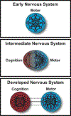
Evolution of motor-cognitive neural linkage. Early brain consisted of a simple sensory-motor system for basic sensing and behaving to a changing environment. Over time, cognitive regions conferred an evolutionary advantage by facilitating abilities such as memory and executive functions, and were built upon existing sensory-motor infrastructure. In the most developed nervous systems (e.g., primates), cognitive and motor regions became specialized and discrete, yet maintained an interdependence.

References

    1. Aboitiz F, Garcia RR, Bosman C, Brunetti E, 2006. Cortical memory mechanisms and language origins. Brain Lang 98, 40–56. DOI: 10.1016/j.bandl.2006.01.006. - DOI - PubMed
    1. Akkal D, Dum RP, Strick PL, 2007. Supplementary motor area and presupplementary motor area: targets of basal ganglia and cerebellar output. The Journal of neuroscience : the official journal of the Society for Neuroscience 27, 10659–10673. - PMC - PubMed
    1. Allen G, McColl R, Barnard H, Ringe WK, Fleckenstein J, Cullum CM, 2005. Magnetic resonance imaging of cerebellar-prefrontal and cerebellar-parietal functional connectivity. Neuroimage 28, 39–48. DOI: 10.1016/j.neuroimage.2005.06.013. - DOI - PubMed
    1. Andersen RA, Essick GK, Siegel RM, 1987. Neurons of Area-7 Activated by Both Visual-Stimuli and Oculomotor Behavior. Experimental Brain Research 67, 316–322. - PubMed
    1. Awh E, Jonides J, Smith EE, Schumacher EH, Koeppe RA, Katz S, 1996. Dissociation of storage and rehearsal in verbal working memory: Evidence from positron emission tomography. Psychological Science 7, 25–31. DOI: DOI 10.1111/j.1467-9280.1996.tb00662.x. - DOI

Publication types

MeSH terms