Review
. 2019 Aug:57:135-144.
doi: 10.1016/j.sbi.2019.03.029. Epub 2019 Apr 28.

Structural changes in the transport cycle of the mitochondrial ADP/ATP carrier

Affiliations
Review

Structural changes in the transport cycle of the mitochondrial ADP/ATP carrier

Jonathan J Ruprecht et al. Curr Opin Struct Biol. 2019 Aug.

Abstract

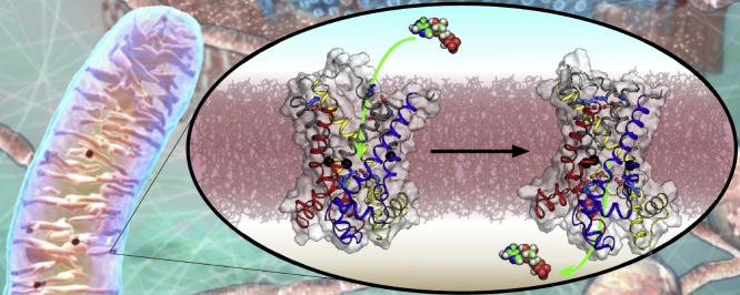
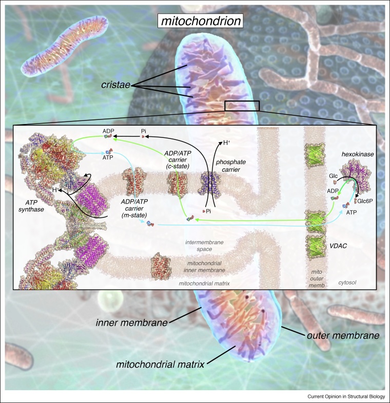
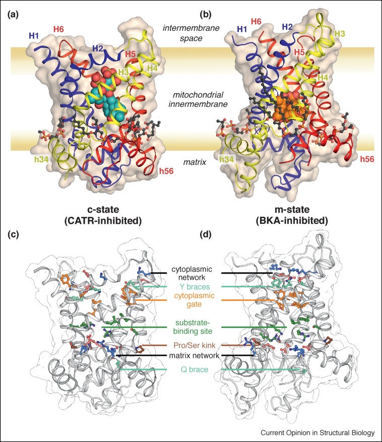
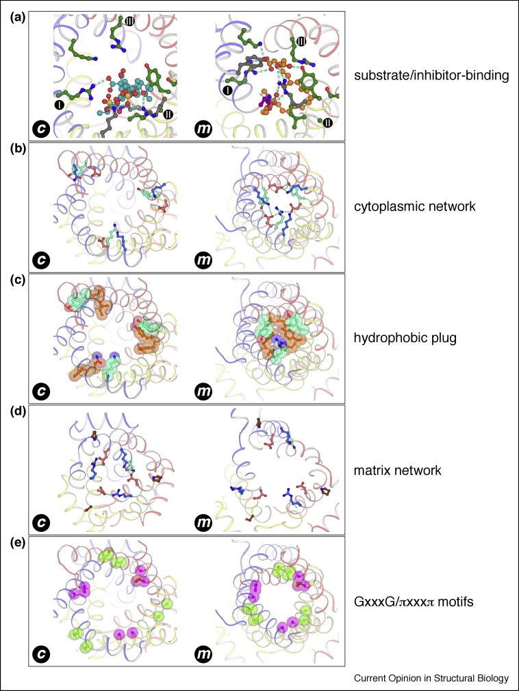
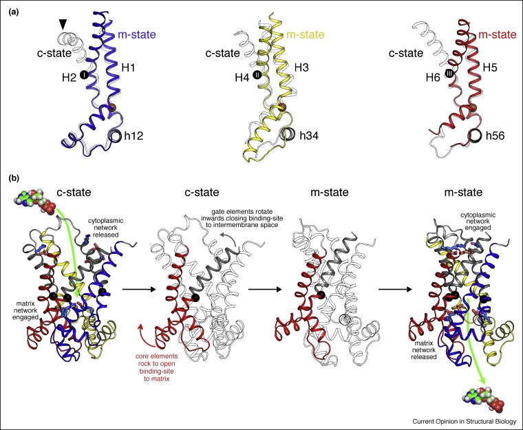
The mitochondrial ADP/ATP carrier, also called adenine nucleotide translocase, accomplishes one of the most important transport activities in eukaryotic cells, importing ADP into the mitochondrial matrix for ATP synthesis, and exporting ATP to fuel cellular activities. In the transport cycle, the carrier changes between a cytoplasmic and matrix state, in which the central substrate binding site is alternately accessible to these compartments. A structure of a cytoplasmic state was known, but recently, a structure of a matrix-state in complex with bongkrekic acid was solved. Comparison of the two states explains the function of highly conserved sequence features and reveals that the transport mechanism is unique, involving the coordinated movement of six dynamic elements around a central translocation pathway.


Figures

None
Graphical abstract
Figure 1
Figure 1
The role of ADP and ATP transport in oxidative phosphorylation. In eukaryotes, the mitochondrion is the site of oxidative phosphorylation. The impermeable mitochondrial inner membrane forms characteristic invaginations, called cristae, and contains the respiratory complexes (not shown), ATP synthase, and transport proteins including the ADP/ATP carrier and phosphate carrier. ATP drives many biochemical reactions (exemplified by hexokinase, PDB: 3B8A, converting glucose to glucose-6-phosphate). The spent fuel ADP crosses the mitochondrial outer membrane via voltage-dependent anion channels (VDAC, PDB: 3EMN) and diffuses into the mitochondrial intermembrane space. ADP binds to mitochondrial ADP/ATP carriers in the c-state (PDB: 4C9H chain A), inducing a conformational change that switches the carrier to the m-state, leading to transport of ADP across the membrane. Phosphate ions are transported across the mitochondrial inner membrane by the phosphate carrier (homology model based upon PDB: 4C9H chain A), in symport with protons. ADP and phosphate are the substrates for ATP synthase (shown in its dimeric form at the sharply curved edge of the crista, PDB: 6B8H). ATP synthase uses the proton motive force generated by the respiratory complexes to drive the rotary mechanism of ATP synthesis [49]. The product, ATP, binds to mitochondrial ADP/ATP carriers in the m-state (PDB: 6GCI chain A), driving a conformational change to the c-state and transport of ATP into the intermembrane space, from where it can diffuse to the cytosol and other cell organelles to fuel more reactions.
Figure 2
Figure 2
Structure of fungal mitochondrial ADP/ATP carriers. (a) The c-state (inhibited by CATR, PDB: 4C9H chain A) and (b) the m-state (inhibited by BKA, PDB: 6GCI chain A). The carrier proteins are shown in cartoon representation, colored by domain (blue, domain 1; yellow, domain 2; red, domain 3), and as a wheat-colored surface. Transmembrane α-helices (H1–H6) and matrix α-helices (h34 and h56, h12 is not labelled) are indicated. The inhibitors are shown in sphere representation with cyan carbons for CATR and orange carbons for BKA. (c) and (d), the same views of the two states, respectively, but with highly conserved sequence features highlighted in color, as indicated.
Figure 3
Figure 3
Key structural elements required for the transport mechanism, observed in the c-state and m-state structures. Domains are represented and colored as in Figure 2. (a) Substrate-binding site residues, colored in green. Also shown are the bound inhibitors CATR (cyan carbons) and BKA (orange carbons). (b) The cytoplasmic network, showing positively charged (blue carbons) and negatively charged residues (red carbons). The tyrosine braces and arginine substitution are shown with cyan carbons. (c) Residues of the hydrophobic plug (orange carbons), which includes the tyrosine brace (cyan carbons). (d) The matrix network, with charged residues shown as in (b). The glutamine brace is shown with cyan carbons. The proline/serine kink residues are shown in brown. BKA has been removed from the view of the m-state structure, for clarity, and E37 has been modelled as a common rotamer. (e) Residues of the GxxxG (magenta carbons) and πxxxπ motifs (green carbons).
Figure 4
Figure 4
Proposed transport mechanism of the mitochondrial ADP/ATP carrier. (a) Conformational changes between c-state (PDB: 4C9H chain A, shown in outline) and m-state (PDB: 6GCI chain A) shown for each domain of the carrier. For the m-state, domains 1–3 are shown as blue, yellow, and red cartoons, respectively. The orange spheres mark the positions of the conserved prolines/serines of the [PS]x[DE]xx[KR] motif. The numbered black spheres mark the positions of the substrate-binding site contact points. Domains have been aligned on their core elements. (b) Conformational changes between modelled uninhibited c-state and m-state of the ADP/ATP carrier, viewed laterally from the membrane. Core elements 1, 2, and 3 are colored by domain in blue, yellow, and red, respectively, and the gate elements are colored grey. Conformational changes between c-state and m-state can be described as a rotation of the core elements, coupled with an inward movement of the gate elements, and are highlighted in the middle panels for domain 3.

References

    1. Kunji E.R.S., Aleksandrova A., King M.S., Majd H., Ashton V.L., Cerson E., Springett R., Kibalchenko M., Tavoulari S., Crichton P.G. The transport mechanism of the mitochondrial ADP/ATP carrier. Biochim Biophys Acta. 2016;1863:2379–2393. - PubMed
    2. Comprehensive review of the structure and function of mitochondrial ADP/ATP carriers, referencing earlier studies.

    1. Garrett R., Grisham C.M. edn 4. Brooks/Cole; Cengage Learning; International ed. Australia; United Kingdom: 2010. Biochemistry.
    1. Buono M.J., Kolkhorst F.W. Estimating ATP resynthesis during a marathon run: a method to introduce metabolism. Physiol Educ. 2001;25:70–71.
    1. Kunji E.R.S. The mitochondrial family of transport proteins. In: Egelman E., editor. vol 8. Elsevier; 2012. pp. 174–205. (Comprehensive Biophysics).
    1. Monne M., Palmieri F. Antiporters of the mitochondrial carrier family. Curr Top Membr. 2014;73:289–320. - PubMed
    2. Review covering other members of the mitochondrial carrier family.

Publication types

MeSH terms

Substances

LinkOut - more resources