

doi: 10.15252/emmm.201810204.
Efficacy of simultaneous VEGF-A/ANG-2 neutralization in suppressing spontaneous choroidal neovascularization
Affiliations
- PMID: 31040126
- PMCID: PMC6505683
- DOI: 10.15252/emmm.201810204
Item in Clipboard
Efficacy of simultaneous VEGF-A/ANG-2 neutralization in suppressing spontaneous choroidal neovascularization
EMBO Mol Med.
2019 May.
Abstract
This study independently confirms in vivo in the
Conflict of interest statement
RF, FR, SU, SG, and CU are employees of F. Hoffmann‐La Roche Ltd.
Figures
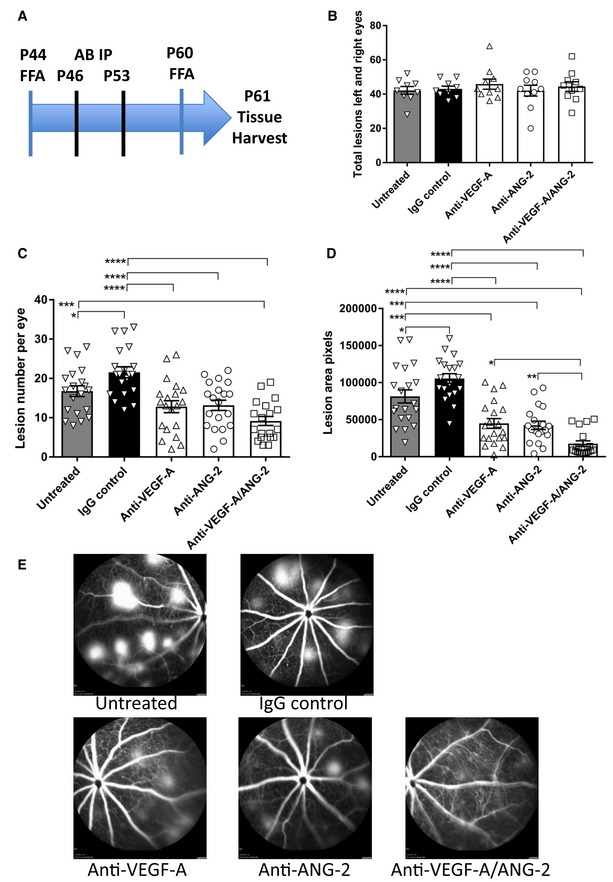
(A) Study schematic. Baseline FFA was carried out at P44‐45. Antibody injections were given IP on P46 and P53. Post‐treatment FFA was at P60 and tissue harvest at P61. (B) Bar/scatter graph of baseline lesion numbers. Lesions were counted in all mice prior to study start. Combined total lesions from left and right eyes were calculated, and then, animals were assigned to treatment groups ensuring no statistically significant differences between groups (P > 0.05). (C, D) Bar/scatter graphs showing numbers of spontaneously occurring lesions (C) and area by fluorescence angiography (D) after two weekly doses of antibody (IgG, anti‐VEGF ‐A, anti‐ANG ‐2 at 5 mg/kg IP , and anti‐VEGF ‐A/ANG ‐2 at 10 mg/kg IP ) followed by analysis a week after the last treatment. (E) Representative examples of fluorescence fundus angiograms from the untreated (top left), IgG control (top middle; 10 mg/kg), anti‐VEGF ‐A (bottom left; 5 mg/kg), anti‐ANG ‐2 (bottom middle; 5 mg/kg), and anti‐VEGF ‐A/ANG ‐2 (bottom right; 10 mg/kg) groups. Data information: SEM is shown as error bars with n = 9–10 animals (B) or n = 19–20 (C, D) eyes per group and significance indicated by asterisks using ANOVA (B: P > 0.05; C: P < 0.0001; D: P < 0.0001, followed by Newman–Keul's multiple comparison test in C, D). In (C), untreated is significantly different versus IgG control (*P < 0.05) and anti‐VEGF ‐A/ANG ‐2 (***P < 0.001). IgG control is significantly different versus anti‐VEGF ‐A (****P < 0.0001), anti‐ANG ‐2 (****P < 0.0001), and anti‐VEGF ‐A/ANG ‐2 (****P < 0.0001). In (D), untreated is significantly different versus IgG control (*P < 0.05), anti‐VEGF ‐A (***P < 0.001), anti‐ANG ‐2 (***P < 0.001), and anti‐VEGF ‐A/ANG ‐2 (****P < 0.0001). IgG control is significantly different versus anti‐VEGF ‐A (****P < 0.0001), anti‐ANG ‐2 (****P < 0.0001), and anti‐VEGF ‐A/ANG ‐2 (****P < 0.0001). Anti‐VEGF ‐A/ANG ‐2 is significantly different versus anti‐VEGF ‐A (*P < 0.05) and anti‐ANG ‐2 (**P < 0.01). FFA , fluorescein fundus angiography; P, post‐natal day; AB , antibody; IP , intraperitoneal; SEM , standard error of the mean; ANOVA , analysis of variance.Source data are available online for this figure.
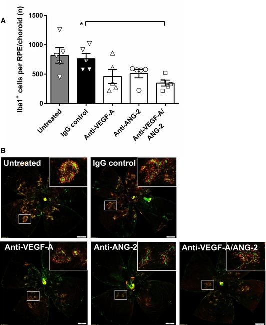
(A) Bar/scatter graph of the number of Iba1+ cells in the subretinal space of mice treated with 10 mg/kg IgG control, 5 mg/kg anti‐VEGF ‐A, 5 mg/kg anti‐ANG ‐2, 10 mg/kg anti‐VEGF ‐A/ANG ‐2, or left untreated. Data are shown as mean ± SEM with n = 5 animals per group, and asterisk denotes significant changes after one‐way ANOVA and Tukey's multiple t‐test (Tukey–Kramer HSD ). Anti‐VEGF ‐A/ANG ‐2 is significantly different from IgG control (*P < 0.02). (B) Representative examples of flat‐mounted RPE /choroid Iba1 (red) and IB 4 counterstain (green) of untreated (top left), IgG control (top middle; 10 mg/kg), anti‐VEGF ‐A (bottom left; 5 mg/kg), anti‐ANG ‐2 (bottom middle; 5 mg/kg), and anti‐VEGF ‐A/ANG ‐2 (bottom right; 10 mg/kg). Adjustment of brightness to 100 using Photoshop CS 6 was applied equally to all images, and non‐tissue background was removed using Lasso tool. Inserts show selected areas. Scale bars = 500 μm. SEM , standard error of the mean; ANOVA , analysis of variance.Source data are available online for this figure.
References
-
- Campochiaro PA, Khanani A, Singer M, Patel S, Boyer D, Dugel P, Kherani S, Withers B, Gambino L, Peters K et al (2016) Enhanced benefit in diabetic macular edema from AKB‐9778 Tie2 activation combined with vascular endothelial growth factor suppression. Ophthalmology 123: 1722–1730 - PubMed
-
- Kienast Y, Klein C, Scheuer W, Raemsch R, Lorenzon E, Bernicke D, Herting F, Yu S, The HH, Martarello L et al (2013) Ang‐2‐VEGF‐A CrossMab, a novel bispecific human IgG1 antibody blocking VEGF‐A and Ang‐2 functions simultaneously, mediates potent antitumor, antiangiogenic, and antimetastatic efficacy. Clin Cancer Res 19: 6730–6740 - PubMed
-
- Liang WC, Wu X, Peale FV, Lee CV, Meng YG, Gutierrez J, Fu L, Malik AK, Gerber HP, Ferrara N et al (2006) Cross‐species vascular endothelial growth factor (VEGF)‐blocking antibodies completely inhibit the growth of human tumor xenografts and measure the contribution of stromal VEGF. J Biol Chem 281: 951–961 - PubMed
Publication types
MeSH terms
Substances
Associated data
- Actions
- Actions
LinkOut - more resources
Full Text Sources
Miscellaneous
