. 2019 Jun 14;294(24):9476-9488.
doi: 10.1074/jbc.RA119.008321. Epub 2019 Apr 30.

Directed evolution of the metalloproteinase inhibitor TIMP-1 reveals that its N- and C-terminal domains cooperate in matrix metalloproteinase recognition

Affiliations

Directed evolution of the metalloproteinase inhibitor TIMP-1 reveals that its N- and C-terminal domains cooperate in matrix metalloproteinase recognition

Maryam Raeeszadeh-Sarmazdeh et al. J Biol Chem. .

Abstract

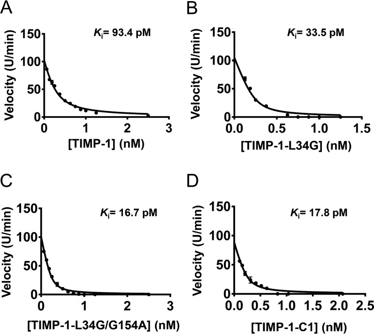
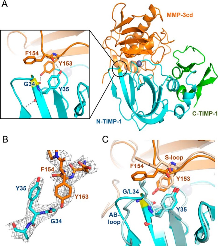
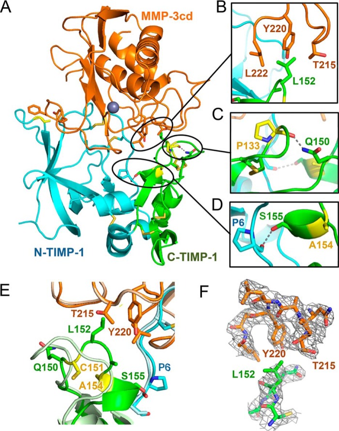
Tissue inhibitors of metalloproteinases (TIMPs) are natural inhibitors of matrix metalloproteinases (MMPs), enzymes that contribute to cancer and many inflammatory and degenerative diseases. The TIMP N-terminal domain binds and inhibits an MMP catalytic domain, but the role of the TIMP C-terminal domain in MMP inhibition is poorly understood. Here, we employed yeast surface display for directed evolution of full-length human TIMP-1 to develop MMP-3-targeting ultrabinders. By simultaneously incorporating diversity into both domains, we identified TIMP-1 variants that were up to 10-fold improved in binding MMP-3 compared with WT TIMP-1, with inhibition constants (Ki ) in the low picomolar range. Analysis of individual and paired mutations from the selected TIMP-1 variants revealed cooperative effects between distant residues located on the N- and C-terminal TIMP domains, positioned on opposite sides of the interaction interface with MMP-3. Crystal structures of MMP-3 complexes with TIMP-1 variants revealed conformational changes in TIMP-1 near the cooperative mutation sites. Affinity was strengthened by cinching of a reciprocal "tyrosine clasp" formed between the N-terminal domain of TIMP-1 and proximal MMP-3 interface and by changes in secondary structure within the TIMP-1 C-terminal domain that stabilize interdomain interactions and improve complementarity to MMP-3. Our protein engineering and structural studies provide critical insight into the cooperative function of TIMP domains and the significance of peripheral TIMP epitopes in MMP recognition. Our findings suggest new strategies to engineer TIMP proteins for therapeutic applications, and our directed evolution approach may also enable exploration of functional domain interactions in other protein systems.

Keywords: crystal structure; directed evolution; matrix metalloproteinase (MMP); metalloprotease; protease inhibitor; protein domain; protein engineering; protein structure; protein-protein interaction; tissue inhibitor of metalloproteinase (TIMP); yeast surface display.


Conflict of interest statement

The authors declare that they have no conflicts of interest with the contents of this article

Figures

Figure 1.
Figure 1.
TIMP-1 yeast display and MMP-3cd binding optimization. A, TIMP-1 yeast surface display constructs in pCHA vector. Top construct (prepro-hTIMP-1), prepro-α-factor yeast signal with Kex2 (KR) and Ste13 (EA) cleavage sites fused to the mature N terminus of TIMP-1 followed by c-myc epitope tag fused to the N terminus of Aga2. Bottom construct (prepro-ΔEA-hTIMP-1), prepro-α-factor yeast signal with Kex2 (KR) cleavage site fused to the mature N terminus of TIMP-1 followed by c-myc epitope tag fused to the N terminus of Aga2. B–D, flow cytometry results for MMP-3cd binding to TIMP-1 constructs displayed on the yeast surface. Left panels, dual scatter plot representing MMP-3cd binding on the x axis and TIMP-1 expression (c-myc binding) on the y axis; Q2 represents the cell population dually labeled by biotinylated MMP-3cd and anti-c-myc binding. Middle panels, histograms of TIMP-1 expression (detected by anti-c-myc). Right panels, histograms of MMP-3cd binding. B, yeast display of human TIMP-1 (hTIMP-1) with Kex2 and Ste13 cleavage sites (prepro-hTIMP-1) shows high expression but only modest MMP-3cd binding, suggesting inefficient processing by Ste13. C, yeast display of human TIMP-1 with Kex2 cleavage site only (prepro-ΔEA-hTIMP-1) shows high expression and MMP-3dc binding, indicating more efficient processing of the mature N terminus of TIMP-1. D, yeast display of human TIMP-1, codon-optimized for yeast, with Kex2 cleavage site only shows expression and MMP-3cd binding similar to the noncodon-optimized construct.
Figure 2.
Figure 2.
Screening a library of TIMP-1 mutants for MMP-3 binding. A, schematic diagram illustrates how a library of TIMP-1 mutants (blue/green) was displayed on the yeast surface. TIMP-1 expression was measured using fluorescent conjugated c-myc antibody (red star), and MMP-3 binding was measured using biotinylated MMP-3cd (orange) and fluorescent conjugated streptavidin (purple star). TIMP-1 variants with improved MMP-3cd binding were screened using FACS. B, library diversity was focused in 17 residues of TIMP-1 (loops in red), located in both the N-terminal (blue) and C-terminal (green) domains, that interact with bound MMP-3cd (orange) in PDB structure 1UEA. Targeted residues are located in the AB-loop, C-connector, and EF-loop of the N-terminal domain, and the GH-loop and MTL of the C-terminal domain. C, the WT TIMP-1 N- and C-terminal domain sequences are shown, colored in blue and green, respectively. Segments that interact with MMP-3cd in crystal structure 1UEA are annotated in black text above the sequence, including the N terminus and AB-loop, C-connector, EF-loop, GH-loop, and MTL. TIMP-1 residues diversified in the targeted library are highlighted in red and underlined. D, flow cytometry scatter plots of dually labeled yeast cells show lower MMP-3cd binding signal (x axis) for naïve library (center panel) relative to WT TIMP-1 (left panel); the population after three rounds of FACS sorting (right panel) shows greatly increased MMP-3 binding signal. P1, the diagonal sort gate, represents a population of yeast cells with a high ratio of MMP-3cd binding relative to TIMP-1 expression.
Figure 3.
Figure 3.
TIMP-1 variants with improved MMP-3 binding. A, flow cytometry scatter plots of dually labeled yeast cells are shown for four yeast-displayed TIMP-1 variants with improved MMP-3cd–binding activity; WT TIMP-1 is shown for reference on the left. The x axis (APC channel) represents biotinylated MMP-3cd binding (250 nm); the y axis (FITC channel) represents TIMP-1 expression. B, median fluorescence MMP-3cd binding signal, corrected for background and normalized to TIMP-1 expression, is plotted for each yeast-displayed TIMP-1 variant stained with 250 nm biotinylated MMP-3cd. Flow cytometry binding experiments were repeated at least twice; plotted values represent average ± S.D. (error bars). C, mutations found in TIMP-1 variants with improved MMP-3 binding are shown, along with their locations in the five targeted MMP-3–interacting loops of TIMP-1 (AB-loop, C-connector (C-conn), EF-loop, GH-loop, and MTL). *, p ≤ 0.05; **, p ≤ 0.01; ***, p ≤ 0.001.
Figure 4.
Figure 4.
Dissecting functionally important mutations of TIMP-1 variants. A, among tested mutations found in the library-selected TIMP-1 variants with improved MMP-3cd–binding affinity, only L34G and T98D show functional enhancement as single mutations. B–D, Gly-154 mutations to Ala, His, or Lys cooperatively enhance MMP-3cd binding when combined with L34G. B, although not function-enhancing as a single mutation, G154A combined with L34G increases MMP-3cd binding nearly to the level of the composite library-selected variant C1 (L34G/L133P/L152C/G154A). C, likewise, G154H has no significant functional effect as a single mutation but cooperatively enhances MMP-3cd binding in combination with L34G, up to the level of the composite library-selected variant C9 (L34G/S68P/L133H/G154H). D, similarly, G154K offers functional benefit only in combination with L34G where it cooperatively enhances MMP-3cd binding nearly to the level of the composite library-selected variant C14 (L34G/T98P/L133N/S134M/G154K). E, by contrast, the G154A mutation does not confer further enhancement of MMP-3cd binding when combined with the T98D mutation. F, the G154H mutation likewise does not significantly improve MMP-3cd binding in the double T98D/G154H variant. Graphs show MMP-3cd binding to TIMP-1 expression ratio, based on median fluorescence corrected to background signal, for yeast-displayed TIMP-1 mutants stained with 250 nm biotinylated MMP-3cd. Flow cytometry binding experiments were repeated at least twice; plotted values represent average ± S.D. (error bars). *, p ≤ 0.05; **, p ≤ 0.01; ns, not significant.
Figure 5.
Figure 5.
Ki determination using Morrison fits of inhibition assays. Equilibrium inhibition constants (Ki) of purified soluble TIMP-1 and variants toward MMP-3cd were measured by the reduction in cleavage rates of a fluorogenic substrate in the presence of increasing concentration of the inhibitors. Data were plotted as initial velocities versus TIMP variant concentration and fitted by multiple regression to Morrison's tight binding inhibition equation as shown. The Ki value determined for each TIMP-1 variant is indicated on the plot and in Table 1. A, WT TIMP-1. B, TIMP-1-L34G. C, TIMP-1-L34G/G154A. D, TIMP-1-C1 (L34G/L133P/L151C/G154A).
Figure 6.
Figure 6.
Crystal structure of TIMP-1-L34G mutant bound to MMP-3cd. A, cartoon representation of the TIMP-1-L34G/MMP-3cd complex crystal structure is shown (right); TIMP-1-L34G is in blue (N-terminal domain) and green (C-terminal domain) with the mutated residue in yellow, and MMP-3cd is in orange with the catalytic zinc ion shown as a gray sphere. The inset panel (left) shows interactions of TIMP-1-L34G Tyr-35 and MMP-3cd Tyr-153, which form a reciprocal tyrosine clasp at the binding interface. B, stick representation of the protein environment surrounding the reciprocal tyrosine clasp with 2FoFc map contoured at 1.5σ. C, superposition of the mutant TIMP-1-L34G/MMP-3cd crystal structure (colored as above) with the WT TIMP-1/MMP-3cd structure (PDB code 1UEA; shown in pale blue/pale orange) highlights the conformational changes in the AB-loop of TIMP-1-L34G, which result in the cinching of the reciprocal tyrosine clasp.
Figure 7.
Figure 7.
Crystal structure of TIMP-1-C1 variant bound to MMP3cd. A, cartoon representation of the TIMP-1-C1/MMP-3cd complex crystal structure is shown using the same color scheme described for Fig. 6. Altered inter- and intramolecular interactions attributable to TIMP-1-C1 mutations are highlighted in B–D. B, a hydrophobic cluster is formed at the intermolecular interface between TIMP-1 Leu-152 and MMP-3cd residues Thr-215, Tyr-220, and Leu-222. C, a new H-bond within the TIMP-1-C1 C-terminal domain is formed between the carbonyl of Pro-133 and the side chain of Gln-150. D, as a consequence of the G154A mutation, residues 154–157 adopt an α-helical conformation, and a new interdomain H-bond is formed between the side chain of Ser-155 in the C-terminal domain and the carbonyl of Pro-6 in the N-terminal domain. E, superposition of the TIMP-1-C1/MMP-3cd crystal structure (colored as above) with the WT TIMP-1/MMP-3cd structure (PDB code 1UEA; shown in pale blue/pale orange) highlights conformational changes in the multiple-turn loop of TIMP-1-C1, which facilitate stabilizing interdomain interactions within TIMP-1 as well as favorable hydrophobic interactions at the interface with MMP-3cd. F, stick representation of the protein environment surrounding the hydrophobic cluster at the intermolecular interface with 2FoFc map contoured at 1.0σ.

References

    1. Huang J., Koide A., Makabe K., and Koide S. (2008) Design of protein function leaps by directed domain interface evolution. Proc. Natl. Acad. Sci. U.S.A. 105, 6578–6583 10.1073/pnas.0801097105 - DOI - PMC - PubMed
    1. Vogel C., Bashton M., Kerrison N. D., Chothia C., and Teichmann S. A. (2004) Structure, function and evolution of multidomain proteins. Curr. Opin. Struct. Biol. 14, 208–216 10.1016/j.sbi.2004.03.011 - DOI - PubMed
    1. Bagowski C. P., Bruins W., and Te Velthuis A. J. (2010) The nature of protein domain evolution: shaping the interaction network. Curr. Genomics 11, 368–376 10.2174/138920210791616725 - DOI - PMC - PubMed
    1. Brew K., and Nagase H. (2010) The tissue inhibitors of metalloproteinases (TIMPs): an ancient family with structural and functional diversity. Biochim. Biophys. Acta 1803, 55–71 10.1016/j.bbamcr.2010.01.003 - DOI - PMC - PubMed
    1. Batra J., and Radisky E. S. (2014) Tissue inhibitors of metalloproteinases (TIMPs): inhibition of Zn-dependent metallopeptidases, in Encyclopedia of Inorganic and Bioinorganic Chemistry (Scott R. A., ed) John Wiley and Sons, Chichester, UK

Publication types

MeSH terms

Substances