. 2019 Apr 30;10(2):e00555-19.
doi: 10.1128/mBio.00555-19.

Binding of Staphylococcus aureus Protein A to von Willebrand Factor Is Regulated by Mechanical Force

Affiliations

Binding of Staphylococcus aureus Protein A to von Willebrand Factor Is Regulated by Mechanical Force

Felipe Viela et al. mBio. .

Abstract

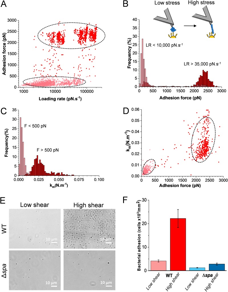
Binding of Staphylococcus aureus to the large plasma glycoprotein von Willebrand factor (vWF) is controlled by hydrodynamic flow conditions. Currently, we know little about the molecular details of this shear-stress-dependent interaction. Using single-molecule atomic force microscopy, we demonstrate that vWF binds to the S. aureus surface protein A (SpA) via a previously undescribed force-sensitive mechanism. We identify an extremely strong SpA-vWF interaction, capable of withstanding forces of ∼2 nN, both in laboratory and in clinically relevant methicillin-resistant S. aureus (MRSA) strains. Strong bonds are activated by mechanical stress, consistent with flow experiments revealing that bacteria adhere in larger amounts to vWF surfaces when the shear rate is increased. We suggest that force-enhanced adhesion may involve conformational changes in vWF. Under force, elongation of vWF may lead to the exposure of a high-affinity cryptic SpA-binding site to which bacteria firmly attach. In addition, force-induced structural changes in the SpA domains may also promote strong, high-affinity binding. This force-regulated interaction might be of medical importance as it may play a role in bacterial adherence to platelets and to damaged blood vessels.IMPORTANCEStaphylococcus aureus protein A (SpA) binds to von Willebrand factor (vWF) under flow. While vWF binding to SpA plays a role in S. aureus adherence to platelets and endothelial cells under shear stress, the molecular basis of this stress-dependent interaction has not yet been elucidated. Here we show that the SpA-vWF interaction is regulated by a new force-dependent mechanism. The results suggest that mechanical extension of vWF may lead to the exposure of a high-affinity cryptic SpA-binding site, consistent with the shear force-controlled functions of vWF. Moreover, strong binding may be promoted by force-induced structural changes in the SpA domains. This study highlights the role of mechanoregulation in controlling the adhesion of S. aureus and shows promise for the design of small inhibitors capable of blocking colonization under high shear stress.

Keywords: Staphylococcus aureus; adhesion; atomic force microscopy; mechanical force; von Willebrand factor.


Figures

FIG 1
FIG 1
Attachment of S. aureus to immobilized vWF and to endothelial cells. (A) Microtiter wells coated with vWF were incubated with S. aureus Newman WT and Newman Δspa bacteria, rinsed, and stained with crystal violet, and the absorbance at 595 nm was measured in an ELISA plate reader. Attachment of WT bacteria was also performed in the presence of 2 μg soluble SpA. (B) Optical microscopy images of S. aureus Newman WT and Newman Δspa bacteria adhering to vWF-coated substrates. (C) vWF release by endothelial cells. Confluent HUVEC monolayers were incubated with increasing concentrations of calcium ionophore A23187 for 10 min. The vWF released in the extracellular matrix was determined with rabbit anti-vWF polyclonal antibody followed by HRP-conjugated goat anti-rabbit. (D) Imaging of vWF release. Shown are confocal microscopy images of HUVECs before (left) and after (right) treatment with calcium ionophore. Staining with mouse anti-vWF antibody and secondary goat anti-mouse Alexa Fluor 647 antibody documents the release of large amounts of vWF upon ionophore treatment (vWF is in red, and the nucleus is in blue). The inset shows a control experiment in which the primary antibody was missing. (E) Endothelial cell adhesion assays. Confluent HUVECs were incubated with S. aureus Newman WT and Newman Δspa bacteria for 90 min, in the presence or absence of 1 μM ionophore. The number of adhering bacteria was determined as described in Materials and Methods. (F) Imaging of bacterial-endothelial cell adhesion. Confluent HUVECs, treated (right) or not (left) with ionophore, were incubated with S. aureus Newman WT bacteria stained with the BacLight viability kit, rinsed, and imaged by confocal microscopy. The arrows indicate bacteria adhering to the endothelial cell surface. For all plots, means and SD of results from two independent experiments, each performed in triplicate, are presented. Statistically significant difference is indicated (Student's t test; *, P < 0.05).
FIG 2
FIG 2
Adhesion forces between S. aureus and vWF in the extracellular matrix. (A) Maximum adhesion force (left) and rupture length (right) histograms with representative retraction force profiles (insets) obtained by recording force-distance curves in HEPES buffer between Newman bacteria and ionophore-treated endothelial cells. Data from a total of 1,536 curves from 3 different bacterial-HUVEC cell pairs are shown. (B) Force data collected between Newman bacteria and nontreated endothelial cells (1,536 curves from 3 cell pairs). (C) Force data collected between Newman Δspa bacteria and ionophore-treated endothelial cells (1,536 curves from 3 cell pairs). The percentage shown represents the proportion of nonadhesive events. For data on more cells, see Table S1.
FIG 3
FIG 3
Adhesion forces between S. aureus and immobilized vWF. (A and B) Maximum adhesion force (left) and rupture length (right) histograms with representative retraction force profiles obtained by recording force distance curves in PBS between Newman WT (A) or Newman Δspa (B) bacteria and vWF-coated substrates. (C and D) Force data obtained for the clinically relevant LAC* strain (C) and its LAC* Δspa mutant (D). For each panel, data from three representative bacterial cells are shown.
FIG 4
FIG 4
Strength of single SpA-vWF bonds. (A) Maximum adhesion force histograms (left) with force maps (insets; image size = 500 nm) and rupture length histograms (right) with representative retraction force profiles (insets) obtained by recording force-distance curves in PBS between 3 different Newman WT bacteria and vWF-modified AFM tips. (B) Data obtained under the same conditions for Newman Δspa bacteria. (Data from 3 cells are merged.)
FIG 5
FIG 5
Mechanical activation of vWF binding to SpA. (A) Strength of single SpA-vWF bonds measured at increasing loading rates (LRs) on Newman WT bacteria (2,468 adhesive events from 6 cells). All adhesion peaks were analyzed to take into account all possible interactions. (B) Analysis of the data in panel A showing that strong bonds are favored at high LR. Discrete ranges of LRs were binned, and the forces were plotted as histograms (see Fig. S3 in the supplemental material). (C) Stiffness of the SpA-vWF complex (km) at low (<500 pN) and high (>500 pN) tensile forces. A cutoff of 500 pN was chosen as essentially all low adhesion forces at low stress were smaller than this value (see panel B). (D) Plot of km as a function of the adhesion force showing that the high mechanical stability of the bond correlates with a high molecular stiffness. (E and F) SpA-dependent bacterial adhesion to vWF increases with fluid shear stress. Shown are optical microscopy images of Newman WT and Δspa bacteria adhering to vWF-coated surfaces in a microparallel flow chamber under low and high shear stresses. Shown in panel F is the quantification of the amounts of adhering bacteria estimated from the experiments described in panel E from a total of 6 images from 3 experiments for each condition.

Comment in

References

    1. Moreillon P, Que Y-A. 2004. Infective endocarditis. Lancet 363:139–149. doi:10.1016/S0140-6736(03)15266-X. - DOI - PubMed
    1. Fowler VG Jr, Miro JM, Hoen B, Cabell CH, Abrutyn E, Rubinstein E, Corey GR, Spelman D, Bradley SF, Barsic B, Pappas PA, Anstrom KJ, Wray D, Fortes CQ, Anguera I, Athan E, Jones P, van der Meer JTM, Elliott TSJ, Levine DP, Bayer AS, ICE Investigators. 2005. Staphylococcus aureus endocarditis: a consequence of medical progress. JAMA 293:3012–3021. doi:10.1001/jama.293.24.3012. - DOI - PubMed
    1. Pappelbaum KI, Gorzelanny C, Grässle S, Suckau J, Laschke MW, Bischoff M, Bauer C, Schorpp-Kistner M, Weidenmaier C, Schneppenheim R, Obser T, Sinha B, Schneider SW. 2013. Ultra-large von Willebrand factor fibers mediate luminal Staphylococcus aureus adhesion to an intact endothelial cell layer under shear stress. Circulation 128:50–59. doi:10.1161/CIRCULATIONAHA.113.002008. - DOI - PubMed
    1. Claes J, Vanassche T, Peetermans M, Liesenborghs L, Vandenbriele C, Vanhoorelbeke K, Missiakas D, Schneewind O, Hoylaerts MF, Heying R, Verhamme P. 2014. Adhesion of Staphylococcus aureus to the vessel wall under flow is mediated by von Willebrand factor-binding protein. Blood 124:1669–1676. doi:10.1182/blood-2014-02-558890. - DOI - PMC - PubMed
    1. Claes J, Liesenborghs L, Peetermans M, Veloso TR, Missiakas D, Schneewind O, Mancini S, Entenza JM, Hoylaerts MF, Heying R, Verhamme P, Vanassche T. 2017. Clumping factor A, von Willebrand factor-binding protein and von Willebrand factor anchor Staphylococcus aureus to the vessel wall. J Thromb Haemost 15:1009–1019. doi:10.1111/jth.13653. - DOI - PMC - PubMed

Publication types

MeSH terms

Substances

LinkOut - more resources