. 2019 Apr 30;12(579):eaau3568.
doi: 10.1126/scisignal.aau3568.

NF-κB signaling dynamics is controlled by a dose-sensing autoregulatory loop

Affiliations

NF-κB signaling dynamics is controlled by a dose-sensing autoregulatory loop

Mialy M DeFelice et al. Sci Signal. .

Abstract

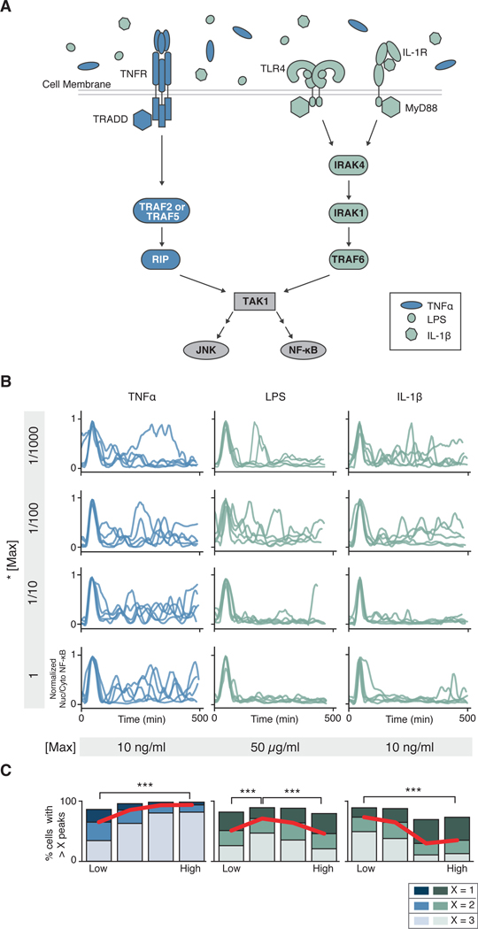
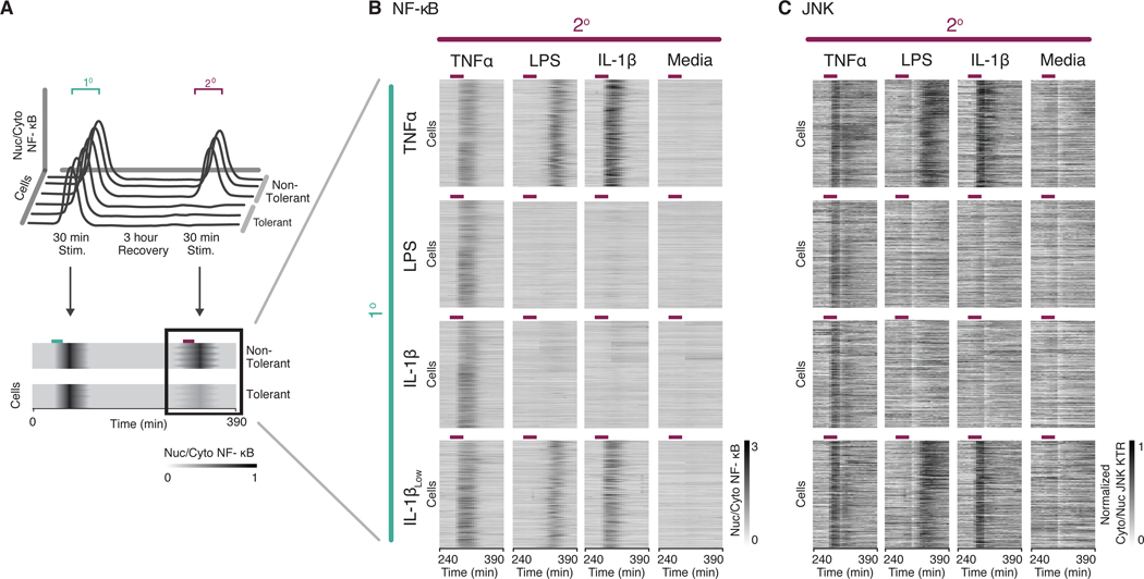
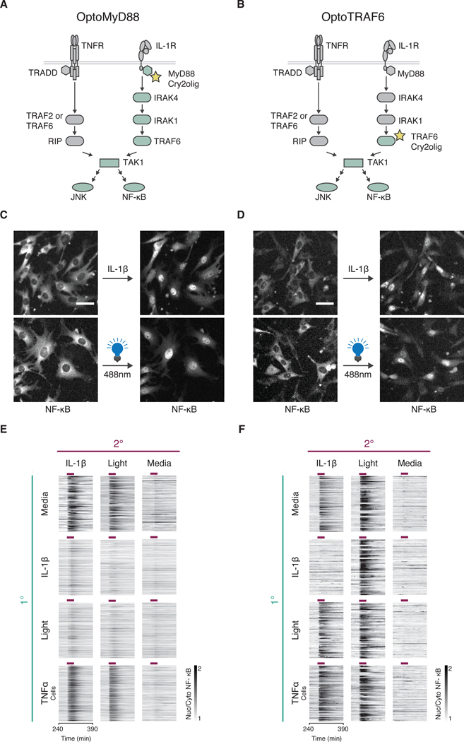
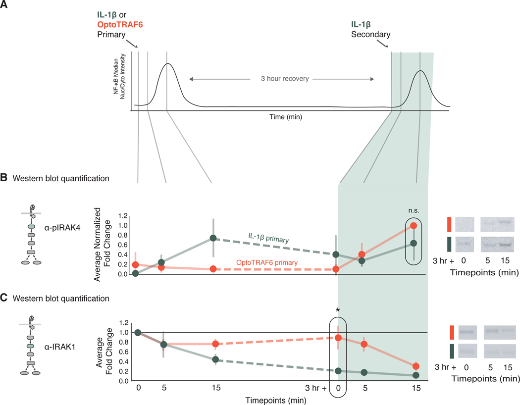
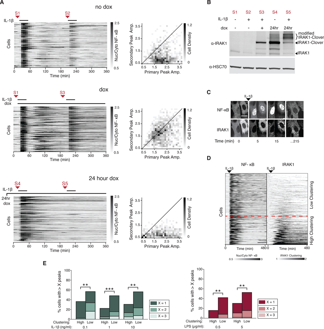
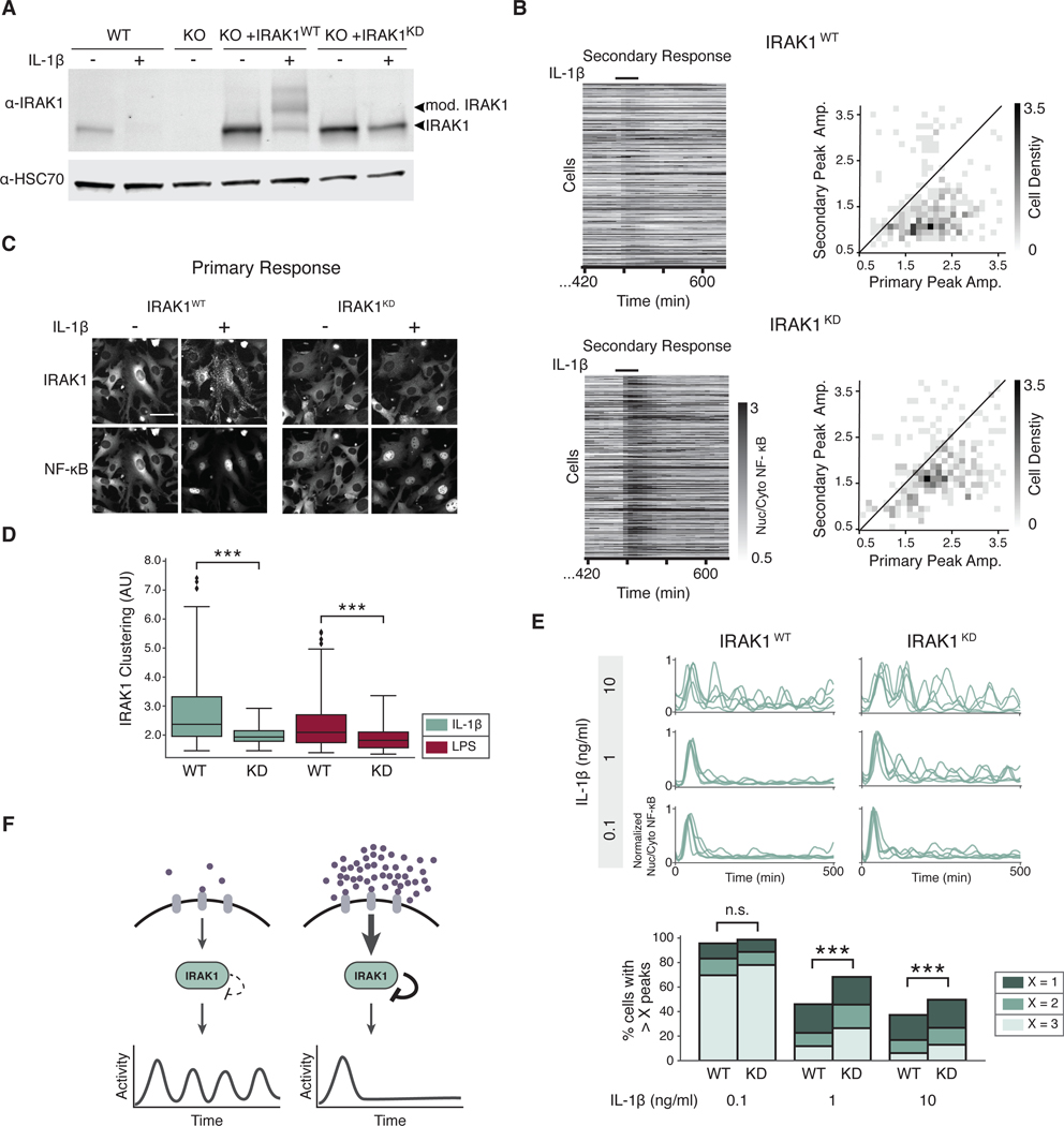
Over the last decade, multiple studies have shown that signaling proteins activated in different temporal patterns, such as oscillatory, transient, and sustained, can result in distinct gene expression patterns or cell fates. However, the molecular events that ensure appropriate stimulus- and dose-dependent dynamics are not often understood and are difficult to investigate. Here, we used single-cell analysis to dissect the mechanisms underlying the stimulus- and dose-encoding patterns in the innate immune signaling network. We found that Toll-like receptor (TLR) and interleukin-1 receptor (IL-1R) signaling dynamics relied on a dose-dependent, autoinhibitory loop that rendered cells refractory to further stimulation. Using inducible gene expression and optogenetics to perturb the network at different levels, we identified IL-1R-associated kinase 1 (IRAK1) as the dose-sensing node responsible for limiting signal flow during the innate immune response. Although the kinase activity of IRAK1 was not required for signal propagation, it played a critical role in inhibiting the nucleocytoplasmic oscillations of the transcription factor NF-κB. Thus, protein activities that may be "dispensable" from a topological perspective can nevertheless be essential in shaping the dynamic response to the external environment.


Figures

Fig. 1.
Fig. 1.. The TNFR and TLR or IL-1R elicit qualitatively different dose-dependent NF-κB dynamics.
(A) Schematic representation of the innate immune signaling network. TNFα, LPS, and IL-1β activate innate immune signaling through different receptors and signaling proteins. While TLR4 (LPS) and IL-1R (IL-1β) share most signaling components, TNFR (TNFα) signals through different proteins. Both cascades lead to activation of NF-κB and MAPK signaling via TAK1. (B) Parental strain (PS) cells (NIH 3T3, RelA−/−, p65-dsRed, H2B-EGFP) were treated with increasing concentrations (top to bottom) of indicated stimuli, imaged, and quantified as described in the Methods (see sections on live cell imaging and segmentation and tracking). Five randomly selected traces from > 2,000 are shown per condition. Traces have been aligned to their first peak for clarity. (C) Peak counting quantification (see Methods for details) of the data presented in (B). Fractions of cells with more than 1, 2, or 3 peaks are shown to highlight population distribution. Data are representative of three independent experiments. High vs. Low, ***P< 0.001 by chi-square test. For TNFα, LPS and IL-1β, > 6,700, 9,600 and 11,600 cells respectively were analyzed across concentration.
Fig. 2.
Fig. 2.. TLR and IL-1R stimuli render cells cross-tolerant to further stimulation.
(A) Schematic representation of the experimental timeline. During tolerance experiments, cells were stimulated with a primary (1°) and secondary (2°) stimulation for 30 minutes with an intervening rest period of 3 hours. Traces of NF-κB activity during the secondary response were converted to heatmaps wherein rows indicate individual cells, columns indicate time, and the grayscale colormap represents nuclear to cytoplasmic median intensity ratio of p65-DsRed. (B and C) Parental Strain (PS) cells (RelA−/−, p65-DsRed, H2B-EGFP) expressing JNK-KTR-mCerulean3 were stimulated with different combinations of primary and secondary inputs as indicated (10 ng/ml TNFα, 5 μg/ml LPS, 1 ng/ml IL-1β, 0.1 ng/ml IL-1βLow), and monitored for NF-κB (B) and JNK (C) activity. Time period for secondary stimulation only is shown for clarity. Purple lines over heatmaps indicate time period when cells were in secondary stimulus. Cells were filtered to include only those responding to the primary stimulus. Data represents two independent experiments (n > 300 cells per condition, > 9,000 cells total).
Fig. 3.
Fig. 3.. Optogenetic control of signaling at MyD88 and TRAF6 nodes maps the cross-tolerance mechanism to IRAK proteins.
(A and B) Schematic representation of OptoMyD88 (A) or OptoTRAF6 (B) activation of innate immune signaling. Upon light stimulation, Opto tools activate their respective downstream proteins labeled in light green. (C and D) Cells (NIH 3T3, p65-mRuby, H2B-iRFP) expressing OptoMyD88 (C) or OptoTRAF6 (D) under the Tet-Responsive Element Third Generation (TRE3G) promoter were incubated in doxycycline (dox, 2 μg/ml) overnight and stimulated with either IL-1β (1 ng/ml) or light (470/24 nm, five 250 ms pulses with 5 min intervals). Representative NF-κB localization images before and after indicated stimuli are shown. Experiment was performed with a 10x objective. Scale bar, 50 μm. (E and F) Secondary response heatmaps of NF-κB nuclear translocation in OptoMyD88 (E) or OptoTRAF6 (F) cells with varied primary (1°) and secondary (2°) stimulations (experimental timeline as in Fig. 2A). Cells were incubated in dox (2 μg/ml) overnight and treated with TNFα (10 ng/ml), IL-1β (1 ng/ml), light (470/24 nm, 250 ms pulses), or media for 30 minutes (purple bars). Data represents three independent experiments (n > 100 cells per condition). Cells were filtered to include only those responding to the primary stimulus, except for those with no primary stimulus.
Fig. 4.
Fig. 4.. Low IRAK1 abundance correlates with the cross-tolerant state.
(A) Schematic detailing of stimulation and sample collection timeline for western blotting. Cells were stimulated with primary stimuli as indicated [1 ng/ml IL-1β, green; OptoTRAF6 (light, 488nm), orange] and sampled at 0, 5, and 15 min after primary stimulation. Cells were washed after 30 min of primary stimulation, allowed to recover for 3 hours, then challenged with secondary stimulation of IL-1β, and sampled at 0, 5 and 15 min. (B) Quantification of immunoblots for phosphorylated IRAK4 (pIRAK4) in cells treated with light (orange) or IL-1β (green) and subjected to a secondary IL-1β stimulation. pIRAK4 abundance was first normalized to a β-actin loading control, then fold-change compared to the unstimulated condition (first time point) was calculated for each sample, then normalized between 0 and 1 across all time points. Data are means ± SD of three independent experiments, n.s. = not significant by a t-test. Representative blots are shown for the last three timepoints, for both IL-1β and OptoTRAF6 primary conditions. Full blots and additional stimulation combinations are presented in fig. S4. (C) Quantification of Western blots for IRAK1, as described in (B). IRAK1 expression was first normalized to a β-actin loading control, then fold-change to the unstimulated condition was calculated for each sample. Data are mean ± SD of three independent experiments, *p<0.05 by a t-test. Representative blots are shown for the last three timepoints, for both IL-1β and OptoTRAF6 primary conditions. As in (B), full blots and additional stimulation combinations are shown in fig. S4.
Fig. 5.
Fig. 5.. Expression of unmodified IRAK1 protein bypasses tolerance.
(A) Induced expression of IRAK1 after primary stimulation. Cells containing TRE3G::IRAK1-Clover were incubated without doxycycline (no dox), with dox at the time of primary stimulation (dox), or 24 hours overnight (24 hour dox). Cells were stimulated with IL-1β (1 ng/ml) and imaged as described in the Methods. After 30 min, cells were washed and allowed to recover for 3 hours and stimulated again with IL-1β (1 ng/ml). Red arrows indicate sample collection points for the Western blot shown in (B). 2D histograms show the distribution of peak amplitudes of nuclear/cytoplasmic NF-κB median intensity during the primary vs secondary response in each cell. Black line indicates primary equals secondary NF-κB amplitude. Data represents three independent experiments with n>100 cells. (B) Western blotting for IRAK1 protein abundance between primary and secondary stimuli in cells treated as in (A) were harvested at indicated times [S1-S5, corresponding to those in A)]. HSC70 was used as a loading control. Blot is representative of three independent experiments. (C) Representative confocal images of IRAK1-Clover cluster formation following NF-kB activation. Cells stably expressing IRAK1-Clover were imaged before and after IL-1β stimulation (1 ng/ml). Scale bar, 50 μm.(D) IRAK1-Clover cells were imaged for 8 hours and stimulated with IL-1β (0.1 ng/ml) 45 min into the time course. NF-κB nuclear/cytoplasmic intensity ratio (left) and IRAK1-Clover clustering dynamics (right) are displayed in tandem. Heatmap rows are ordered top to bottom based on increasing IRAK1 clustering (see Materials and Methods). Dashed red line represents an arbitrary IRAK1 clustering threshold (1.3-fold change). (E) Grouping of cells by IRAK1 clustering separates oscillatory vs. non-oscillatory cells. Irak1-KO cells expressing IRAK1-Clover were stimulated with IL-1β (0.1, 1, and 10 ng/ml) or LPS (0.5 and 5 μg/ml). Peaks of NF-κB activity and IRAK1 clustering from single cell traces obtained in (D) and fig. S6B were measured as described in the Methods. An arbitrary threshold of 1.3-fold increase in IRAK1 clustering was used in all conditions to group high vs low IRAK1 clustering cells. Within each group, fractions of cells with more than 1, 2, or 3 peaks are shown to highlight population distribution. (n >100 cells; **P<0.01 and ***P<0.001 by chi-square test). Additional clustering quantification is provided in fig. S8A, and heatmaps of additional concentrations of IL-1β are provided in fig. S6B.
Fig. 6.
Fig. 6.. IRAK1 kinase activity is critical to regulate NF-κB signaling dynamics.
(A) WT cells and Irak1-KO cells reconstituted with IRAK1WT or IRAK1KD were incubated with or without IL-1β (1 ng/ml) for 3 hours. Lysate was collected and immunoblotted against IRAK1. Arrows indicate posttranslationally modified and unmodified IRAK1 protein. HSC70 was used as a loading control. Blot is representative of 3 experiments. (B) Secondary response heatmaps of IRAK1WT or IRAK1KD cells show reduced tolerance in IRAK1KD. Irak1-KO cells reconstituted with IRAK1WT and IRAK1KD were stimulated with a 30-min pulse of IL-1β (1 ng/ml), washed, allowed to recover for 8 hours, and stimulated again with a secondary pulse of IL-1β (1 ng/ml). 2D histograms show the distribution of peak amplitudes of nuclear/cytoplasmic NF-κB median intensity during the primary vs. secondary response in each cell. Black line indicates primary equals secondary NF-κB amplitude. Data represents three independent experiments with n>100 cells. (C) Irak1-KO cells expressing IRAK1WT-Clover or IRAK1KD-Clover were imaged prior to and 20 min after stimulation with IL-1β (1 ng/ml). Representative images are shown. Scale bar, 50 μm. (D) IRAK1 clustering was quantified as described in methods in IRAK1WT-Clover or IRAK1KD-Clover cells stimulated with IL-1β (0.1 ng/ml) or LPS (0.5 μg/ml). Data represent n >100 cells; ***P<0.001 by a Kolmogorov–Smirnov test. (E) IRAK1 kinase activity regulates oscillatory dynamics. PS cells expressing IRAK1WT-Clover or IRAK1KD -Clover were stimulated with IL-1β (0.1, 1, or 10 ng/ml) and imaged for 8 hours. Five randomly selected single cell traces are presented for each condition. Peak counting of NF-κB oscillations were counted as described in methods. Fractions of cells with more than 1, 2, or 3 peaks are shown to highlight population distribution. Data represents three independent experiments with n >100 cells (n.s.= not significant; ***p < 0.001; chi-square test). See fig. S8B for TNFα data. (F) Schematic model of the effects of IRAK1-dependent autoinhibitory loop in NF-κB signaling dynamics. When ligand is in low abundance, TLR and IL-1R signaling is not inhibited after the initial activation, and continues to signal in an oscillatory pattern. When ligand concentration is high, IRAK1 kinase activity strongly inhibits signaling following the initial activation, and oscillations are not detected.

References

    1. Kholodenko BN, Cell-signalling dynamics in time and space. Nature reviews. Molecular cell biology 7, 165–176 (2006); published online EpubMar ( 10.1038/nrm1838). - DOI - PMC - PubMed
    1. Purvis JE, Lahav G, Encoding and decoding cellular information through signaling dynamics. Cell 152, 945–956 (2013); published online EpubFeb 28 ( 10.1016/j.cell.2013.02.005). - DOI - PMC - PubMed
    1. Albeck JG, Mills GB, Brugge JS, Frequency-modulated pulses of ERK activity transmit quantitative proliferation signals. Molecular cell 49, 249–261 (2013); published online EpubJan 24 ( 10.1016/j.molcel.2012.11.002). - DOI - PMC - PubMed
    1. Shankaran H, Ippolito DL, Chrisler WB, Resat H, Bollinger N, Opresko LK, Wiley HS, Rapid and sustained nuclear-cytoplasmic ERK oscillations induced by epidermal growth factor. Molecular systems biology 5, 332 (2009) 10.1038/msb.2009.90). - DOI - PMC - PubMed
    1. Sparta B, Pargett M, Minguet M, Distor K, Bell G, Albeck JG, Receptor Level Mechanisms Are Required for Epidermal Growth Factor (EGF)-stimulated Extracellular Signal-regulated Kinase (ERK) Activity Pulses. The Journal of biological chemistry 290, 24784–24792 (2015); published online EpubOct 09 ( 10.1074/jbc.M115.662247). - DOI - PMC - PubMed

Publication types

MeSH terms

LinkOut - more resources