. 2019 Apr 30;9(1):6680.
doi: 10.1038/s41598-019-43051-7.

Single molecule super-resolution imaging of bacterial cell pole proteins with high-throughput quantitative analysis pipeline

Affiliations

Single molecule super-resolution imaging of bacterial cell pole proteins with high-throughput quantitative analysis pipeline

Ipek Altinoglu et al. Sci Rep. .

Abstract

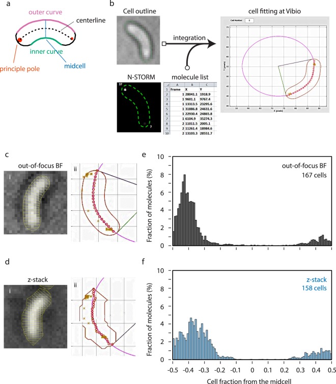
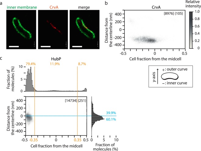
Bacteria show sophisticated control of their cellular organization, and many bacteria deploy different polar landmark proteins to organize the cell pole. Super-resolution microscopy, such as Photo-Activated Localization Microscopy (PALM), provides the nanoscale localization of molecules and is crucial for better understanding of organization and dynamics in single-molecule. However, analytical tools are not fully available yet, in particular for bacterial cell biology. For example, quantitative and statistical analyses of subcellular localization with multiple cells from multiple fields of view are lacking. Furthermore, brightfield images are not sufficient to get accurate contours of small and low contrast bacterial cells, compared to subpixel presentation of target molecules. Here we describe a novel analytic tool for PALM which integrates precisely drawn cell outlines, of either inner membrane or periplasm, labelled by PALM-compatible fluorescent protein fusions, with molecule data for >10,000 molecules from >100 cells by fitting each cell into an oval arc. In the vibrioid bacterium Vibrio cholerae, the polar anchor HubP constitutes a big polar complex which includes multiple proteins involved in chemotaxis and the flagellum. With this pipeline, HubP is shown to be slightly skewed towards the inner curvature side of the cell, while its interaction partners showed rather loose polar localization.


Conflict of interest statement

The authors declare no competing interests.

Figures

Figure 1
Figure 1
Polar HubP clusters. (a,b) Representative image of cell with native level (a) or overexpressed (b) HubP-FPs. Corresponding out-of-focus BF image (i), conventional fluorescent image (ii) are also shown. The region in the purple square is magnified in (iii). Bar = 500 nm. (c–f) Distribution of HubP clusters in native level expression (c and d) or overexpressed (e,f) conditions. (c,e) Dot plots of number of molecules per cluster. For ≥2 clusters per cell, the cluster with highest number of molecules was indicated in red and other clusters were shown in blue. The mean and standard error of mean are also indicated. (d,f) Number of cells containing 1, 2, or 3 clusters of HubP molecules with respect to cell size. 1.28 µm is the average cell size for these experiments.
Figure 2
Figure 2
Vibio, high-throughput quantitative PALM molecule localization analysis pipeline. (a) Schematics of V. cholerae cell. The pole with more detected molecule is determined as principle pole. (b) Graphical abstract of Vibio pipeline. (c,d) (i) Representative cell of single out-of-focus BF image (c) or reconstructed image from 32 z-stacks (d). Outline and segmentation are shown by a yellow line. (ii) Fitting of corresponding cell in Vibio. Each detected HubP molecule is plotted with ‘x’. (e,f) Histogram presentation of the localization of HubP molecules relative to the long axis of the cell determined by indicated outlining method. −0.5 indicates the principle pole and +0.5 indicates the other pole.
Figure 3
Figure 3
Novel cell outlining with photo-activatable/switchable fluorephores. (a) PALM of V. cholerae cells simultaneously expressing ssDsbA-PAmCherry and DronPA-MTS. Representative images reconstructed by N-STORM are shown. (b,c) Representative Images of HubP-DronPA (shown in green) with PAmCherry-MTS (b) or ssDsbA-PAmCherry (c) (shown in red). Out-of-focus BF image (i), conventional fluorescent images of HubP (ii), and outlining molecule (iii) are also shown. (d,e) Localization of HubP molecules relative to the cell length determined by the indicated outlining method. For better comparison, BF outlining results are recapitulated from Fig. 2e. Bar = 1 µm.
Figure 4
Figure 4
Fine-scale localization analyses by Vibio. (a) Representative processed PALM images of V. cholerae cell expressing DronPA-MTS (green) and CrvA-PAmCherry (red). Bar = 1 µm. (b) 2D plot presentation of CrvA-PAmCherry molecule localization. Cell outline was determined by DronPA-MTS. (c) Precise subcellular localization of HubP-DronPA molecules. Cell outline was determined by PAmCherry-MTS. 2D plot and histograms are shown. Percentage of molecules localized in specific fractions (polar, middle cell body, outer side, inner side) are indicated. Total number of molecules detected and number of cells analyzed are shown in square brackets and in braces, respectively.
Figure 5
Figure 5
Fine-scale localization analyses of polar proteins. 2D plot and histogram presentations of DronPA fusions of FlhG (a), FlhF (b), ParC (c,d) and ParP (e,f) in hubP+ (ac,e) or ∆hubP (d,f) background. PAmCherry-MTS was co-expressed for the determination of cell contour. Results are presented as described in Fig. 4c.

References

    1. Surovtsev IV, Jacobs-Wagner C. Subcellular Organization: A Critical Feature of Bacterial Cell Replication. Cell. 2018;172:1271–1293. doi: 10.1016/j.cell.2018.01.014. - DOI - PMC - PubMed
    1. Davis BM, Waldor MK. Establishing polar identity in gram-negative rods. Curr Opin Microbiol. 2013;16:752–759. doi: 10.1016/j.mib.2013.08.006. - DOI - PMC - PubMed
    1. Lenarcic R, et al. Localisation of DivIVA by targeting to negatively curved membranes. EMBO J. 2009;28:2272–2282. doi: 10.1038/emboj.2009.129. - DOI - PMC - PubMed
    1. Ramamurthi KS, Losick R. Negative membrane curvature as a cue for subcellular localization of a bacterial protein. Proc Natl Acad Sci USA. 2009;106:13541–13545. doi: 10.1073/pnas.0906851106. - DOI - PMC - PubMed
    1. Ben-Yehuda S, Rudner DZ, Losick R. RacA, a bacterial protein that anchors chromosomes to the cell poles. Science. 2003;299:532–536. doi: 10.1126/science.1079914. - DOI - PubMed

Publication types

MeSH terms

Substances