Review
. 2019 May-Jun;32(3):264-272.
doi: 10.20524/aog.2019.0355. Epub 2019 Jan 23.

Pathophysiology of internal hemorrhoids

Affiliations
Review

Pathophysiology of internal hemorrhoids

Nikolaos Margetis. Ann Gastroenterol. 2019 May-Jun.

Abstract

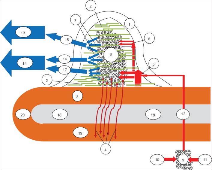
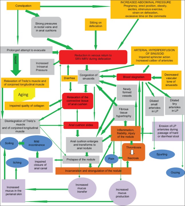
Hemorrhoidal disease is a fairly common and debilitating clinical entity. Despite centuries' of attempts to shed light on its pathophysiology, to cure those affected and to improve sufferers' quality of life, many aspects of the disease remain elusive. Individual beliefs and historical legends, accompanied by undocumented theories, have established and perpetuated the confusion regarding the mechanisms leading to the development of the disease and the rules governing its treatment. Hemorrhoids are classified as internal or external and are viewed as a disease when they become symptomatic. Returning to basic medical sciences, this mini-review focuses on internal hemorrhoids and aims to define the histology and anatomy of the normal and abnormal internal hemorrhoidal plexus and to encourage clinicians to comprehend the pathophysiology of the disease. If doctors can understand the pathophysiology of hemorrhoidal disease, they will be able to clarify the nature of the associated symptoms and complications and to make the correct therapeutic decision.

Keywords: Internal hemorrhoids; anal cushions; anal nodules; prolapse; sliding anal canal.


Conflict of interest statement

Conflict of Interest: None

Figures

Figure 1
Figure 1
A proposed schematic representation of the anatomy of the anal cushion. Anal cushion consists of a non-vascular part (the transitional epithelium – 1, the submucosal connective tissue – 2 and the muscle of Treitz, which has 2 elements, the anal submucosal muscle – 3 and the mucosal suspensory ligament or Park’s ligament - 4) and a vascular part (the small arterioles - 5, the tiny arterioles – 6, the tiny venules – 7 and the direct arteriolar-venular anastomoses between arterioles and venules, that form a subcutaneous spongy network of capillaries, the sinusoids – 8). Arterial blood from the retrorectal plexus - 9 (formed by the SRA -10 and MRA -11) supplies the sinusoidal plexus via 3 terminal arterial branches (here one of them is shown - 12). Before turning into sinusoids - 8, the terminal branch subdivides into small - 5 and tiny - 6 arterioles in the lamina propria. These small and tiny arterioles are the source of bleeding, when the anal cushion is transformed to an anal nodule. Sinusoid drainage is accomplished via tiny venules - 7, which direct venous blood both to the portal circulation – 13 (through the SRV -15) and to the systemic circulation – 14 (mainly through the MRV-16 and to a lesser extent through the IRV -17). The cluster of sinusoids is tightly fixed to the inner anal sphincter -18 by the anal submucosal muscle -3 and by the submucosal connective tissue - 2 (which extends between the sinusoidal anastomoses) and to the conjoined longitudinal muscle -19 by the mucosal suspensory ligament -4. According to several researchers, anal submucosal muscle -3 is thought to represent the continuity of the conjoined longitudinal muscle -19; the latter is believed to form a loop -20 at the lower end of the internal anal sphincter (based on references 5,7,8,10,12-20) IRV, inferior rectal vein; MRA, middle rectal artery; MRV, middle rectal vein; SRA, superior rectal artery; SRV, superior rectal vein
Figure 2
Figure 2
A suggested integrated algorithm summarizing contemporary thinking regarding the pathophysiology of internal hemorrhoids. Hemorrhoidal disease can arise from separate origins or from various combinations of them: these are the eight starting points, shown in the orange boxes (increased intraabdominal pressure, sitting on defecation, constipation, diarrhea, arterial hyperperfusion of the hemorrhoidal plexus, decreased vascular tone of sinusoids, impaired quality of collagen and relaxation of the supporting muscles of the internal hemorrhoidal plexus). These initiation points force the physiological and histological status of the internal hemorrhoidal plexus towards distinct, divergent, abnormal directions. Nonetheless, they finally converge on 4 central pathophysiological events that are almost invariably encountered in internal hemorrhoids. These events (sliding of the anal cushion, relaxation of the connective tissue of the cushion, reduction of venous return from sinusoids to SRV and MRV during defecation and stagnation of blood inside the dilated plexus) are illustrated in the red boxes. The bond between the starting points and the core pathophysiological events is strong and consists of a plethora of consecutive pathophysiological stages, indicated by the gray boxes, which are interconnected, interdependent and inter-fortified. The conceptual network of events leading to hemorrhoidal disease is complicated and has not been fully elucidated in every detail; nevertheless it is established and perpetuated with the assistance of many vicious cycles, either direct—for example between the increased intra-anal pressure and the reduction of venous return during defecation—or multi-step, as between the congestion of sinusoids and incarceration. It is noteworthy that this multimodal and multidirectional network is continuously auto-reinforced, as is implied by the universally green color of the arrows. This means that, according to this model, internal hemorrhoids offer only one option: to become worse over time. Ultimately, symptoms (shown in blue) occur. This happens when the order of events meets critical pathophysiological steps (shown in purple); the erosion of arterioles of the lamina propria of the anal nodule during passage of hard or diarrheal stool is typically responsible for the various forms of hemorrhoidal bleeding. Last, pain, a non-typical symptom of internal hemorrhoids, appears when complications (brown boxes) such as inflammation, incarceration, thrombosis and necrosis arise (based on references 4,7-11,13,15-17,19,2131,34,38,39) LP, lamina propria; MRV, middle rectal vein; SRV, superior rectal vein

References

    1. Holley CJ. History of hemorrhoidal surgery. South Med J. 1946;39:536–541. - PubMed
    1. Senagore AJ. Surgical management of hemorrhoids. J Gastrointest Surg. 2002;6:295–298. - PubMed
    1. Ellesmore S, Windsor AC. Surgical history of haemorrhoids. In: Charles MV, editor. Surgical treatment of haemorrhoids. London: Springer; 2002. pp. 1–4.
    1. Hancock BD. Internal sphincter and the nature of haemorrhoids. Gut. 1977;18:651–655. - PMC - PubMed
    1. Thomson WH. The nature of haemorrhoids. Br J Surg. 1975;62:542–552. - PubMed

LinkOut - more resources