

Phagocytosis in the Brain: Homeostasis and Disease
- PMID: 31040847
- PMCID: PMC6477030
- DOI: 10.3389/fimmu.2019.00790
Phagocytosis in the Brain: Homeostasis and Disease
Erratum in
-
Corrigendum: Phagocytosis in the Brain: Homeostasis and Disease.Front Immunol. 2019 Jul 10;10:1575. doi: 10.3389/fimmu.2019.01575. eCollection 2019. Front Immunol. 2019. PMID: 31354724 Free PMC article.
Abstract
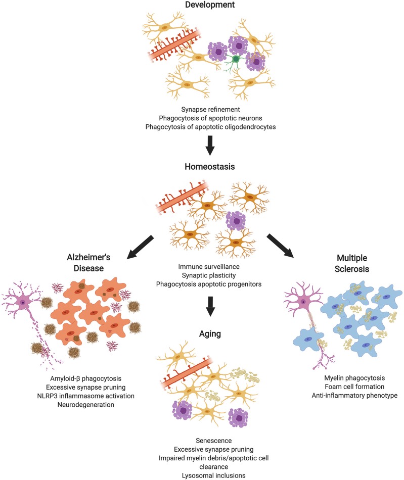
Microglia are resident macrophages of the central nervous system and significantly contribute to overall brain function by participating in phagocytosis during development, homeostasis, and diseased states. Phagocytosis is a highly complex process that is specialized for the uptake and removal of opsonized and non-opsonized targets, such as pathogens, apoptotic cells, and cellular debris. While the role of phagocytosis in mediating classical innate and adaptive immune responses has been known for decades, it is now appreciated that phagocytosis is also critical throughout early neural development, homeostasis, and initiating repair mechanisms. As such, modulating phagocytic processes has provided unexplored avenues with the intent of developing novel therapeutics that promote repair and regeneration in the CNS. Here, we review the functional consequences that phagocytosis plays in both the healthy and diseased CNS, and summarize how phagocytosis contributes to overall pathophysiological mechanisms involved in brain injury and repair.
Keywords: macrophage; microglia; neurodegeneration; neuroinflammation; phagocytosis.
Figures
References
Publication types
MeSH terms
Grants and funding
LinkOut - more resources
Full Text Sources
Other Literature Sources