. 2015 Sep 29;6(3):67-71.
doi: 10.1002/cld.496. eCollection 2015 Sep.

Noncirrhotic portal hypertension: Imaging, hemodynamics, and endovascular therapy

Affiliations

Noncirrhotic portal hypertension: Imaging, hemodynamics, and endovascular therapy

Venkatesh P Krishnasamy et al. Clin Liver Dis (Hoboken). .
No abstract available


Figures

Figure 1
Figure 1
Digital subtraction angiography (DSA) image demonstrating patent TIPS (white thin arrow) connecting the right portal vein (black thin arrow) to the right hepatic vein (black thick arrow) through the liver parenchyma. Embolization coils are present in the coronary vein (white thick arrow) due to persistent reflux into varices.
Figure 2
Figure 2
DSA image demonstrating patent DIPS (white thin arrow) connecting the portal vein (black thin arrow) to the inferior vena cava (IVC) (white thick arrow) through the caudate lobe of the liver. Also visualized is the proximal splenic vein (black thick arrow) and superior mesenteric vein (SMV) (white bent arrow).
Figure 3
Figure 3
(A) Coronal contrast‐enhanced CT image demonstrating a chronically occluded portal vein (white thick arrow) with cavernous transformation (white thin arrow). The superior mesenteric vein (black thick arrow) is widely patent. (B) DSA image demonstrating a patent mesocaval shunt (white thin arrow) connecting the superior mesenteric vein (black thin arrow) to the IVC (white thick arrow). A pigtail flush catheter is in place in the superior mesenteric vein.
Figure 4
Figure 4
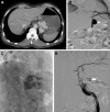
(A) Axial contrast‐enhanced computed tomography (CT) image demonstrating marked gastric varices (white arrow). (B) DSA with balloon occlusion retrograde venography of the gastrorenal shunt (GRS) (white thin arrow) demonstrates opacification of gastric varices (white thick arrow) and the inferior phrenic vein (black thin arrow). The GRS is catheterized via the left renal vein from the IVC. (C) Fluoroscopic image with balloon occlusion retrograde venography demonstrates opacification of the gastric varices (white thin arrows). (D) DSA from GRS (white thin arrow) after embolization of the gastric varices with sclerosant and coils (white thick arrow) demonstrates persistent opacification of the inferior phrenic vein (black thin arrow).

References

    1. Köklü S, Çoban Ş, Yüksel O, Arhan M. Left‐sided portal hypertension. Dig Dis Sci 2007;52:1141‐1149. - PubMed
    1. Khanna R, Sarin SK. Non‐cirrhotic portal hypertension ‐ Diagnosis and management. J Hepatol 2014;60:421‐441. - PubMed
    1. Weltin G, Taylor KJ, Carter AR, Taylor CR. Duplex Doppler: identification of cavernous transformation of the portal vein. AJR Am J Roentgenol 1985;144:999‐1001. - PubMed
    1. Sharma MP, Dasarathy S, Misra SC, Saksena S, Sundaram KR. Sonographic signs in portal hypertension: a multivariate analysis. Trop Gastroenterol 1996;17:23‐29. - PubMed
    1. Maruyama H, Shimada T, Ishibashi H, Takahashi M, Kamesaki H, Yokosuka O. Delayed periportal enhancement: a characteristic finding on contrast ultrasound in idiopathic portal hypertension. Hepatol Int 2012;6:511‐519. - PubMed