

Temporal shifts in the mycobiome structure and network architecture associated with a rat (Rattus norvegicus) deep partial-thickness cutaneous burn
- PMID: 31041451
- PMCID: PMC6939685
- DOI: 10.1093/mmy/myz030
Temporal shifts in the mycobiome structure and network architecture associated with a rat (Rattus norvegicus) deep partial-thickness cutaneous burn
Abstract
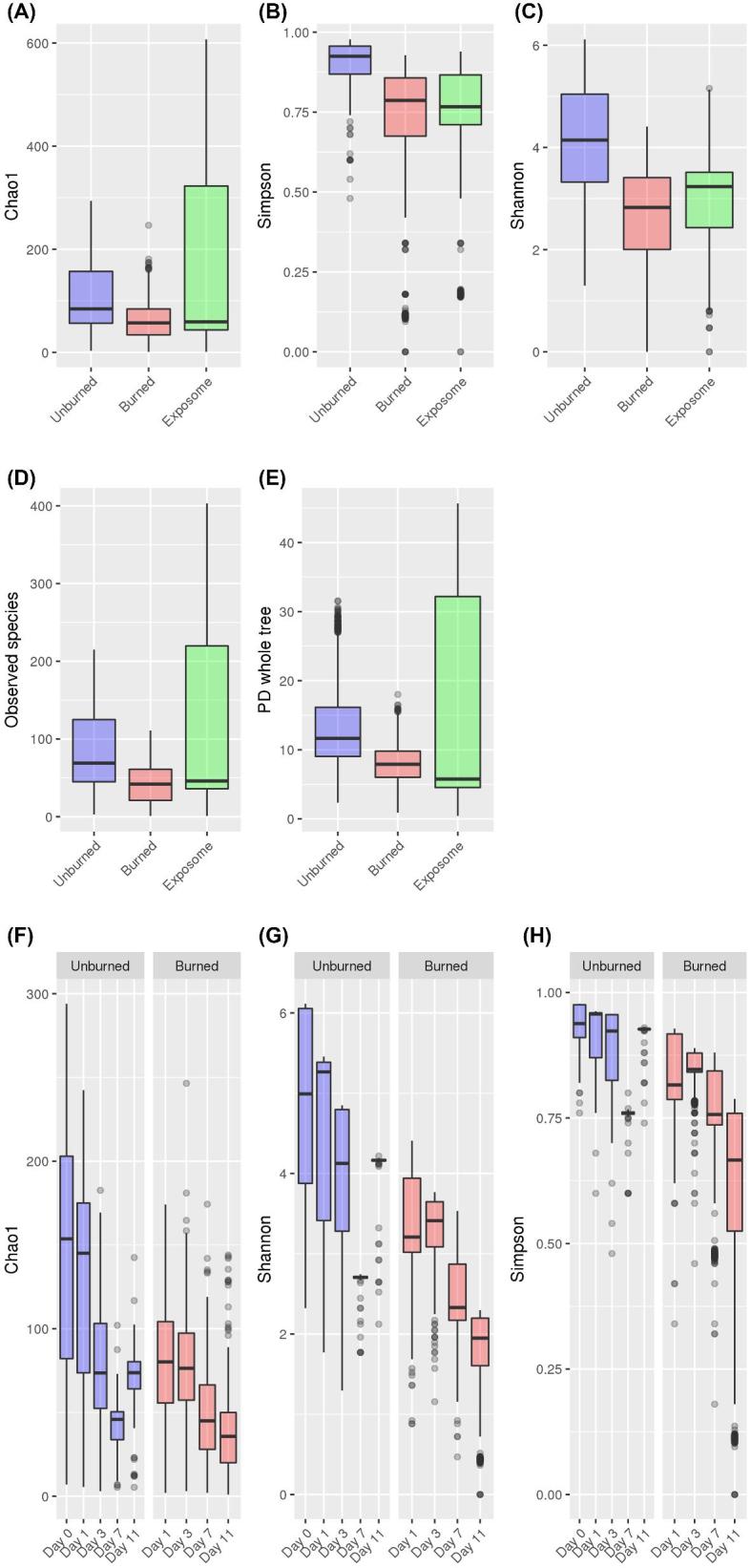
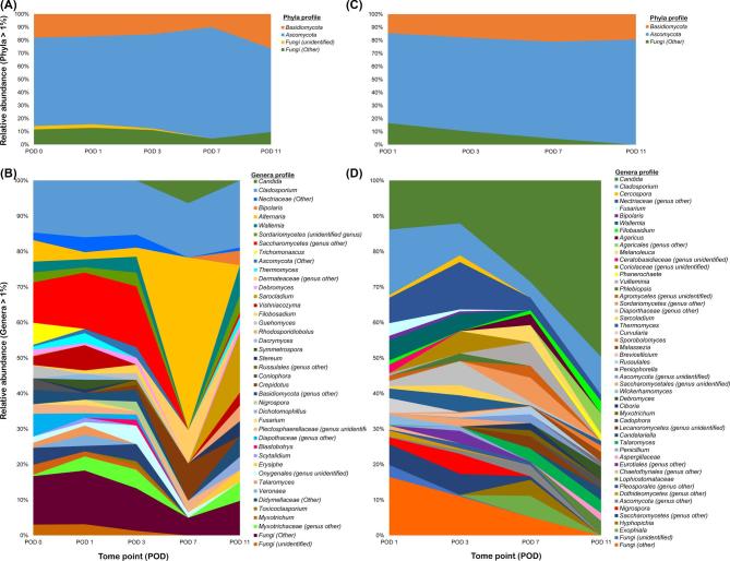
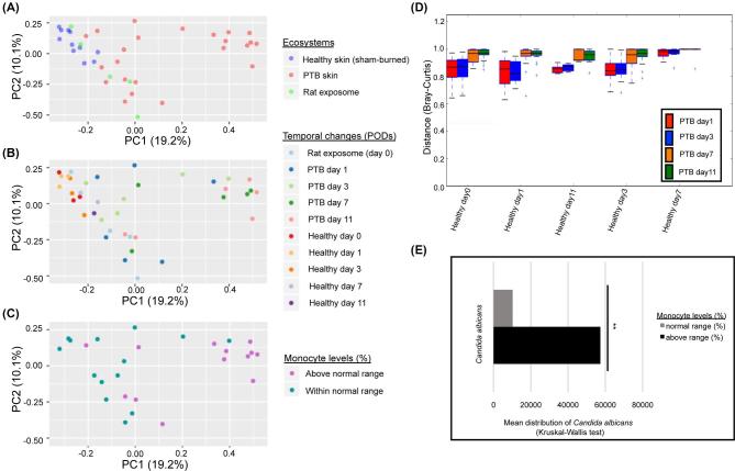
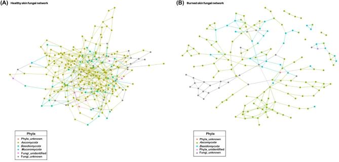
With a diverse physiological interface to colonize, mammalian skin is the first line of defense against pathogen invasion and harbors a consortium of microbes integral in maintenance of epithelial barrier function and disease prevention. While the dynamic roles of skin bacterial residents are expansively studied, contributions of fungal constituents, the mycobiome, are largely overlooked. As a result, their influence during skin injury, such as disruption of skin integrity in burn injury and impairment of host immune defense system, is not clearly delineated. Burn patients experience a high risk of developing hard-to-treat fungal infections in comparison to other hospitalized patients. To discern the changes in the mycobiome profile and network assembly during cutaneous burn-injury, a rat scald burn model was used to survey the mycobiome in healthy (n = 30) (sham-burned) and burned (n = 24) skin over an 11-day period. The healthy skin demonstrated inter-animal heterogeneity over time, while the burned skin mycobiome transitioned toward a temporally stabile community with declining inter-animal variation starting at day 3 post-burn injury. Driven primarily by a significant increase in relative abundance of Candida, fungal species richness and abundance of the burned skin decreased, especially in days 7 and 11 post-burn. The network architecture of rat skin mycobiome displayed community reorganization toward increased network fragility and decreased stability compared to the healthy rat skin fungal network. This study provides the first account of the dynamic diversity observed in the rat skin mycobiome composition, structure, and network assembly associated with postcutaneous burn injury.
Keywords: Rattus norvegicus; burned skin mycobiome; deep-partial thickness burn; rat skin mycobiome; skin fungal community structure; skin fungal network assembly.
Published by Oxford University Press on behalf of The International Society for Human and Animal Mycology 2019.
Figures
Similar articles
-
Identification of Metagenomics Structure and Function Associated With Temporal Changes in Rat (Rattus norvegicus) Skin Microbiome During Health and Cutaneous Burn.J Burn Care Res. 2020 Feb 19;41(2):347-358. doi: 10.1093/jbcr/irz165. J Burn Care Res. 2020. PMID: 31665423
-
Topographical and physiological differences of the skin mycobiome in health and disease.Virulence. 2017 Apr 3;8(3):324-333. doi: 10.1080/21505594.2016.1249093. Epub 2016 Oct 18. Virulence. 2017. PMID: 27754756 Free PMC article. Review.
-
The gut mycobiome of the Human Microbiome Project healthy cohort.Microbiome. 2017 Nov 25;5(1):153. doi: 10.1186/s40168-017-0373-4. Microbiome. 2017. PMID: 29178920 Free PMC article.
-
The vaginal mycobiome: A contemporary perspective on fungi in women's health and diseases.Virulence. 2017 Apr 3;8(3):342-351. doi: 10.1080/21505594.2016.1237332. Epub 2016 Sep 22. Virulence. 2017. PMID: 27657355 Free PMC article. Review.
-
Development of Pseudomonas aeruginosa Biofilms in Partial-Thickness Burn Wounds Using a Sprague-Dawley Rat Model.J Burn Care Res. 2019 Jan 1;40(1):44-57. doi: 10.1093/jbcr/iry043. J Burn Care Res. 2019. PMID: 30137429 Free PMC article.
Cited by
-
Differences in fungal communities in the fur of two- and three-toed sloths revealed by ITS metabarcoding.Microbiology (Reading). 2023 Feb;169(2):001309. doi: 10.1099/mic.0.001309. Microbiology (Reading). 2023. PMID: 36848210 Free PMC article.
-
Colonizing microbiota is associated with clinical outcomes in diabetic wound healing.Adv Drug Deliv Rev. 2023 Mar;194:114727. doi: 10.1016/j.addr.2023.114727. Epub 2023 Feb 8. Adv Drug Deliv Rev. 2023. PMID: 36758858 Free PMC article. Review.
-
Bacterial Interactions in the Context of Chronic Wound Biofilm: A Review.Microorganisms. 2022 Jul 25;10(8):1500. doi: 10.3390/microorganisms10081500. Microorganisms. 2022. PMID: 35893558 Free PMC article. Review.
-
Formation of Pseudomonas aeruginosa Biofilms in Full-thickness Scald Burn Wounds in Rats.Sci Rep. 2019 Sep 20;9(1):13627. doi: 10.1038/s41598-019-50003-8. Sci Rep. 2019. PMID: 31541159 Free PMC article.
References
MeSH terms
Grants and funding
LinkOut - more resources
Full Text Sources
Medical
Research Materials
