. 2019 Dec;234(12):21925-21936.
doi: 10.1002/jcp.28756. Epub 2019 May 1.

The Misshapen subfamily of Ste20 kinases regulate proliferation in the aging mammalian intestinal epithelium

Affiliations

The Misshapen subfamily of Ste20 kinases regulate proliferation in the aging mammalian intestinal epithelium

Qi Li et al. J Cell Physiol. 2019 Dec.

Abstract

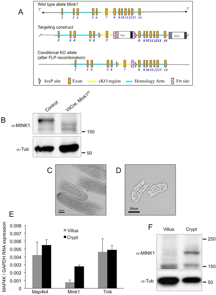
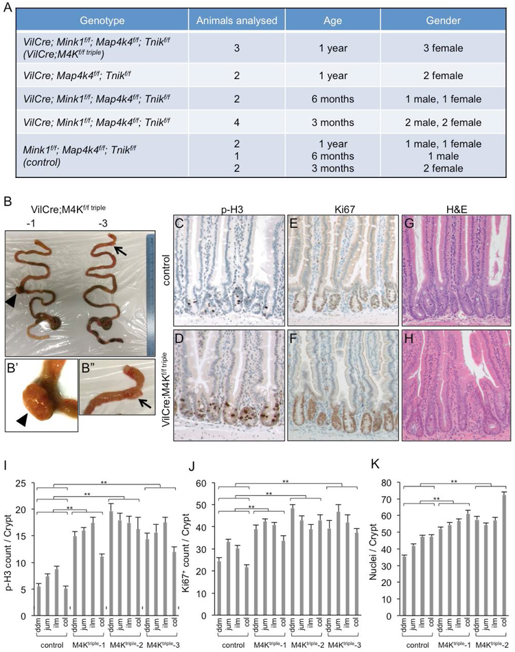
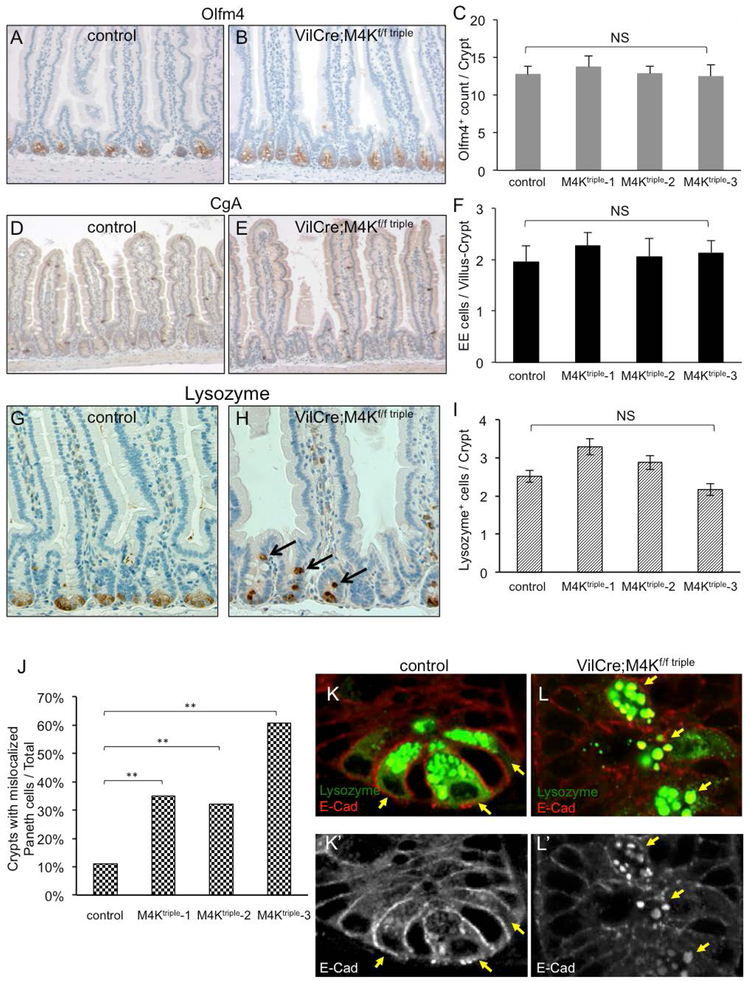
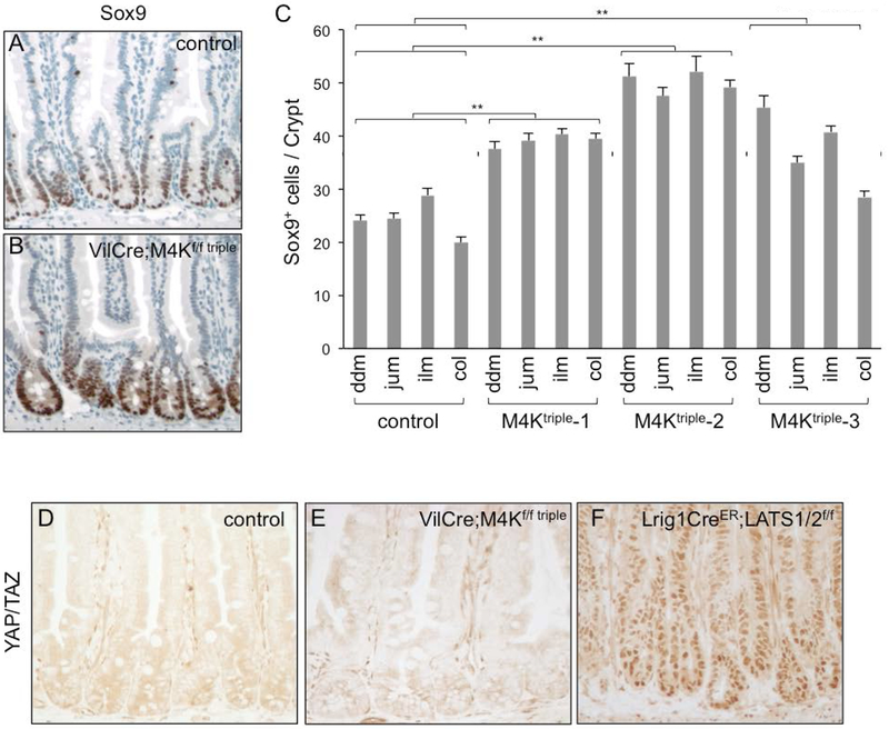
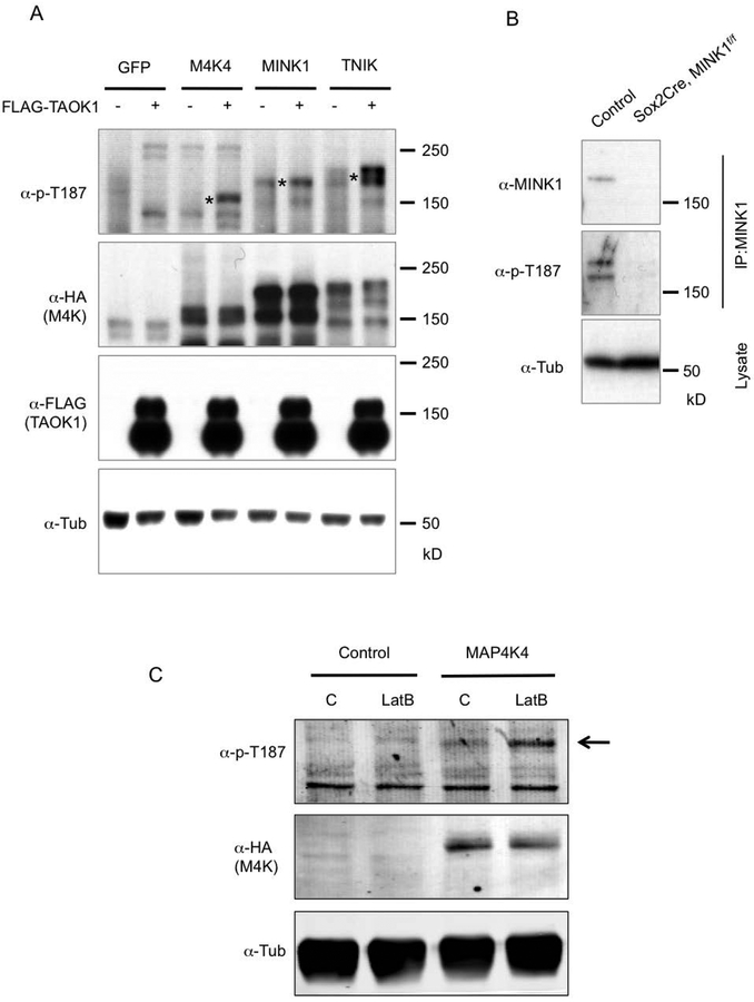
The intestinal epithelium has a high rate of cell turn over and is an excellent system to study stem cell-mediated tissue homeostasis. The Misshapen subfamily of the Ste20 kinases in mammals consists of misshapen like kinase 1 (MINK1), mitogen-activated protein kinase kinase kinase kinase 4 (MAP4K4), and TRAF2 and NCK interacting kinase (TNIK). Recent reports suggest that this subfamily has a novel function equal to the Hippo/MST subfamily as upstream kinases for Warts/Large tumor suppressor kinase (LATS) to suppress tissue growth. To study the in vivo functions of Mink1, Map4k4, and Tnik, we generated a compound knockout of these three genes in the mouse intestinal epithelium. The intestinal epithelia of the mutant animals were phenotypically normal up to approximately 12 months. The older animals then exhibited mildly increased proliferation throughout the lower GI tract. We also observed that the normally spatially organized Paneth cells in the crypt base became dispersed. The expression of one of the YAP pathway target genes Sox9 was increased while other target genes including CTGF did not show a significant change. Therefore, the Misshapen and Hippo subfamilies may have highly redundant functions to regulate growth in the intestinal epithelium, as illustrated in recent tissue culture models.

Keywords: MAP4K4; MINK1; Ste20 kinases; TNIK; hippo; intestine; misshapen.


Figures

Figure 1
Figure 1
The generation of Mink1f/f conditional knockout mouse. (A) The genomic locus of Mink1 is shown in the top panel. The Mink1 conditional knockout mouse strategy, targeting the deletion of exon 7, and the various sites are shown in the panels as indicated. (B) The Western blot showing the endogenous MINK1 protein in intestinal epithelial extracts, and the signal disappeared in the extracts from VilCre;Mink1f/f animals. The anti-Tubulin blot was used as an internal loading control. (C) Phase contrast microscope images of isolated small intestinal villi, which were used in the following quantitative real-time PCR and Western blot. (D) Phase contrast microscope images of similarly isolated small intestinal crypts. (E) Relative mRNA expression for Map4k4, Mink1 and Tnik in the separated villi and crypts. The expression level was normalized to that of GAPDH in parallel qPCR assays. (F) Endogenous protein expression of MINK1 is enriched in intestinal crypts. Extract lysates of indicated tissues were used for Western blot using the antibodies as indicated.
Figure 2
Figure 2
Loss of Mink1, Map4k4 and Tnik leads to increased proliferation in the intestines of aging animals. (A) A list of animals with the indicated genotype, gender and age used for quantification of intestinal phenotypes. The triple knockout animals, abbreviated as VilCre;M4Kf/f triple, showed the proliferation increase only after 1 year. The animals of the same triple knockout genotype at 3 or 6 months, or other combinations, did not show an increase. They had the same proliferation marker measurement as the control animals, which were the triple-floxed littermates without VilCre. (B) Photographs of whole intestines from two VilCre;M4Kf/f triple mice of 1 year old. The small tissue outgrowth of the small intestines are indicated by the arrowhead and arrow, and shown as the enlarged images in B’ and B”. (C-H) Images of immunohistochemical staining in small intestines of 1 year old control and VilCre;M4Kf/f triple mice. Phosphorylated histone 3 (p-H3) stains for mitotic chromosomes. Ki67 is a cell proliferating marker. Hematoxylin and eosin (H&E) shows the morphology to aid the counting of cell number in the intestinal crypts; this staining is included in all these panels, and is shown as double staining in panels C-F and as single staining in panels G-H. (I-K) Quantification of p-H3+, Ki67+ and cell number in control and VilCre;M4Kf/f triple mouse intestines. The counting of positive cell staining per crypt was performed separately in the different regions of the lower GI tract. ddm is duodenum, jum is jejunum, ilm is ileum, col is colon. Three different VilCre;M4Kf/f triple knockout animals (−1, −2, −3) are shown separately. p value indicates the comparison of the data sets in the specific region of the mutant to the same region of the control. All the individual comparisons show statistical significance. Experiments were performed at least 3 times independently. The error bar is standard error of the mean, and p-value is from Student’s T-test: * is p<0.05, ** is p<0.01, NS is no significance with p>0.05.
Figure 3
Figure 3
The MAP4K triple mutant intestines have normal cell fate but mislocalized paneth cells. (A-B) Immunohistochemical staining of the stem cell marker Olfm4, shown as brown staining at the crypt base, together with H&E staining to illustrate the tissue and cell morphology. A control and a triple mutant intestinal tissue are shown. (C) The quantification of the Olfm4+ cell staining in control and mutant intestines is shown. NS is no significance, with P value >0.05. (D-E) The images show immunohistochemical staining of the enteroendocrine cell marker Chromogranin A (CgA) in a control and a triple mutant intestines. (F) The quantification of the CgA+ cell staining in control and mutant intestines is shown. (G-H) The images show immunohistochemical staining of the Paneth cells marker Lysozyme in a control and a triple mutant intestine. (I) The quantification of the Lysozyme+ cell staining in control and mutant intestines is shown. (J) The percentage of crypts with mislocalized Paneth cells from control and triple mutant intestines is shown. For each crypt we examined, if there was one or more Lysozyme+ cell located above the crypt base, as those shown in panel H indicated by arrows, that crypt would be counted as positive for mislocalization. p-values are calculated by applying χ2-test. (K-L) Images of immunofluorescence staining of Lysozyme in green and E-Cadherin (E-Cad) in red, in small intestinal crypts of a control and a triple mutant. The bottom panels show single channel of E-Cad staining of the same images in white. The arrows point to Paneth cells, marked with high level of Lysozyme staining. These cells are localized at the crypt base of control, but are mislocalized into upper part of the crypt of mutant. The E-Cad staining in Paneth cells of the mutant (panel L’) shows lower membrane staining near cell-cell junctions but higher cytoplasmic staining that colocalizes with Lysozyme staining.
Figure 4
Figure 4
Assessment of YAP activity after loss of Mink1, Map4k4 and Tnik. (A-C) The images are immunohistochemical staining using an anti-Sox9 antibody, together with H&E staining. Sox9 nuclear staining shown in brown is strong in crypt nuclei, and appears to be stronger in the triple mutant. Panel C is quantification of the number of cells that are Sox9+ in control and mutant crypts. The increase of Sox9+ cell number in the mutants may result from a combination of increased cell number and increased staining. ** is P value <0.01. (D-F) Images of immunohistochemical staining using an anti-YAP/TAZ antibody in control and mutant intestines. There is no significant nuclear staining even after loss of the three kinases. Meanwhile, the nuclear staining is clear using this antibody after loss of LATS½.
Figure 5
Figure 5
Regulation of MINK1, MAP4K4 and TNIK activities. (A) Western blots using extracts of transfected HEK293 cells. The plasmids containing cDNA of the genes indicated at the top of the panels were transfected into the cells. After 48 hours the cells were used for extract preparation, and the proteins resolved on SDS PAGE and analysed by Western blots using the antibodies indicated to the left of the panels. The asterisks in the top panel indicate the bands matching the predicted sizes of the full length proteins expressed. The MAP4K proteins are tagged with HA at the N-termini. (B) Western blot analysis of endogenous MINK1 protein phosphorylation. Protein extracts were prepared from mouse small intestinal epithelia of control (Mink1f/f without Cre) and mutant, which was 2 months old Sox2Cre; MINK1f/f. An anti-TNIK/MINK1 (BD Transduction Laboratories) antibody was used for IP, and then a rabbit anti-MINK1 (Bethyl) or the anti-p-T187 was used for blots. (C) Western blot analysis for the response of MAP4K4 to change of mechanical property. HEK293 cells were transfected with GFP cDNA as control or MAP4K4 cDNA. After 48 hours, the cells were treated with actin polymerization inhibitor Latrunculin B (LatB) for 1 hour. The cells were then used immediately for lysate preparation. The lysates were used for SDS PAGE and for blots using the indicated antibodies.

Similar articles

Cited by

References

    1. Anazi S, Shamseldin HE, AlNaqeb D, Abouelhoda M, Monies D, Salih MA, Al-Rubeaan K, and Alkuraya FS. 2016. A null mutation in TNIK defines a novel locus for intellectual disability. Hum Genet. 135:773–778. - PubMed
    1. Bankaitis ED, Ha A, Kuo CJ, and Magness ST. 2018. Reserve Stem Cells in Intestinal Homeostasis and Injury. Gastroenterology. 155:1348–1361. - PMC - PubMed
    1. Beumer J, and Clevers H. 2016. Regulation and plasticity of intestinal stem cells during homeostasis and regeneration. Development. 143:3639–3649. - PubMed
    1. Brinkman AS, Murali SG, Hitt S, Solverson PM, Holst JJ, and Ney DM. 2012. Enteral nutrients potentiate glucagon-like peptide-2 action and reduce dependence on parenteral nutrition in a rat model of human intestinal failure. Am J Physiol Gastrointest Liver Physiol. 303:G610–622. - PMC - PubMed
    1. Burette AC, Phend KD, Burette S, Lin Q, Liang M, Foltz G, Taylor N, Wang Q, Brandon NJ, Bates B, Ehlers MD, and Weinberg RJ. 2015. Organization of TNIK in dendritic spines. J Comp Neurol. 523:1913–1924. - PMC - PubMed

Publication types