Review
. 2019 Aug 1;317(2):C167-C176.
doi: 10.1152/ajpcell.00509.2018. Epub 2019 May 1.

Diaphragm contractile weakness due to reduced mechanical loading: role of titin

Affiliations
Review

Diaphragm contractile weakness due to reduced mechanical loading: role of titin

Robbert J van der Pijl et al. Am J Physiol Cell Physiol. .

Abstract

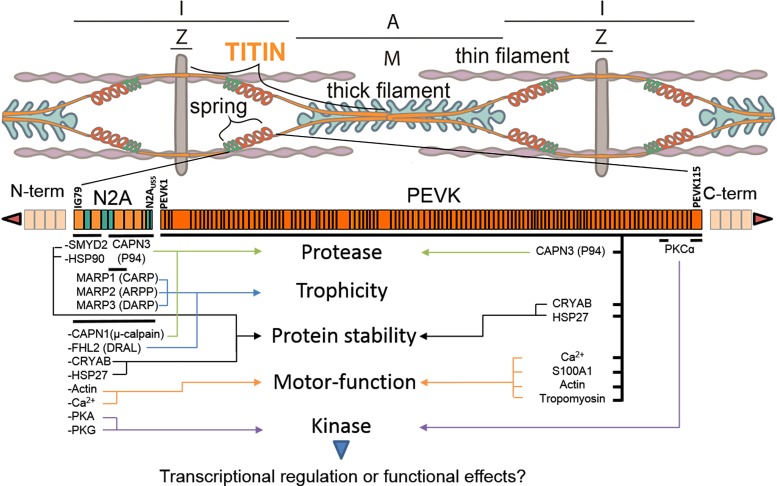
The diaphragm, the main muscle of inspiration, is constantly subjected to mechanical loading. Only during controlled mechanical ventilation, as occurs during thoracic surgery and in the intensive care unit, is mechanical loading of the diaphragm arrested. Animal studies indicate that the diaphragm is highly sensitive to unloading, causing rapid muscle fiber atrophy and contractile weakness; unloading-induced diaphragm atrophy and contractile weakness have been suggested to contribute to the difficulties in weaning patients from ventilator support. The molecular triggers that initiate the rapid unloading atrophy of the diaphragm are not well understood, although proteolytic pathways and oxidative signaling have been shown to be involved. Mechanical stress is known to play an important role in the maintenance of muscle mass. Within the muscle's sarcomere, titin is considered to play an important role in the stress-response machinery. Titin is a giant protein that acts as a mechanosensor regulating muscle protein expression in a sarcomere strain-dependent fashion. Thus titin is an attractive candidate for sensing the sudden mechanical arrest of the diaphragm when patients are mechanically ventilated, leading to changes in muscle protein expression. Here, we provide a novel perspective on how titin and its biomechanical sensing and signaling might be involved in the development of mechanical unloading-induced diaphragm weakness.

Keywords: diaphragm; loading; mechanical ventilation; titin.


Conflict of interest statement

No conflicts of interest, financial or otherwise, are declared by the authors.

Figures

Fig. 1.
Fig. 1.
Schematic illustration of changes in diaphragm loading and their potential effect on muscle trophicity. Left: various mechanical insults to the sarcomere resulting in stress signaling. Center and right: response to mechanical insult resulting in longitudinal (middle) and cross-sectional (right) atrophy or hypertrophy. Note that eccentric contractions are not discussed in the review, but they might occur during asynchrony of the ventilator cycle and spontaneous diaphragm activity in the patient. Gray: Z disks; purple: thin filaments; green; thick filaments; orange: titin. Dashed lines/boxes show the normal state of sarcomere length or muscle (fiber) size. Costal width and thickness are based on human diaphragm at end-expiration. UDD, unilateral diaphragm denervation; PEEP, positive end-expiratory pressure; MV, mechanical ventilation.
Fig. 2.
Fig. 2.
Model of the sarcomere, the smallest contractile unit of striated muscle. The sarcomere comprises 3 major filaments: the thin (mostly actin) filaments, the thick (mostly myosin) filaments, and the giant filamentous molecule titin. The thin filaments are anchored in the Z disk, where they are cross linked by α-actinin. The thick filaments are centrally located in the sarcomere and constitute the sarcomeric A band. The myosin heads, or cross bridges, on the thick filament interact with actin during activation. Titin spans the half-sarcomeric distance from the Z disk to the M band, thus forming a third sarcomeric filament. In the I-band region, titin is extensible and functions as a molecular spring that develops passive tension upon stretch. In the A band, titin is less extensible due to its strong interaction with the thick filament. Titin contains 3 “hot spots” for titin-based sensing and signaling, one at the Z disk, one at the M line, and one at the I band. The hot spot at the I band is shown, with the green spring denoting the N2A segment (domain IG79–N2AUS5) and the orange spring denoting the PEVK region (region rich in proline, glutamic acid, valine, and lysine; domain PEVK1–PEVK115). The accentuated segment shows the domain structure of the spring region in titin, with the rectangles denoting the relative size of the numerous titin domains that encompass the spring (immunoglobulin-like in light orange, unique sequences in green and the PEVK domains in dark orange). Known binding proteins involved in muscle trophicity bind titin in the I-band region forming a titin-specific signaling hub. Signaling proteins are shown with their approximate binding location on titin and their established function in the sarcomere. Ca2+, calcium; CAPN1 (μ-calpain), calpain 1 or μ-calpain; CAPN3 (P94), calpain 3, previously called p94; CRYAB, ɑβ-crystalin; FHL2 and DRAL), four and a half LIM domains protein 2 or downregulated in rhabdomyosarcoma LIM protein; HSP27, heat shock protein 27 (Hsp27), also known as heat shock protein-β1 (HSPB1); HSP90, heat shock protein 90; MARP1 (CARP), muscle ankyrin repeat protein 1 or cardiac ankyrin repeat protein; MARP2 (ARPP), muscle ankyrin repeat protein 2 or ankyrin repeat, PEST sequence, and proline-rich region; MARP3 (DARP), muscle ankyrin repeat protein 3 or diabetes-related ankyrin repeat protein; PKA, protein kinase A; PKCɑ, protein kinase C-α; PKG, protein kinase G; S100A1, S100 calcium-binding protein A1; SMYD2, SET and MYND domain-containing protein 2.
Fig. 3.
Fig. 3.
Diagram of passive-cyclic stretch induced hypertrophy of the diaphragm. A: illustration of unilateral diaphragm denervation (UDD), showing the top (thoracic) view of the diaphragm, consisting of 2 costals attached to the ribcage and the 2 crus leaves attached to the spine, forming a parachute-like muscle group with radial-aligned muscle fibers meeting at the central tendon. Arrows show the main principles of UDD with 1 denervated (right) costal, by transection of 1 of the 2 phrenic nerves [the motor nerves powering the diaphragm, each innervating a single costal and crus leaf (i.e., hemidiaphragm)], and an antiparallel innervated/active (left) costal stretching the denervated costal. The innervated costal shortens ~25%, consistent with a sarcomere working range of ~2.2–2.9 μm (based on a resting length (i.e., end expiration) of 2.9 µm, as assessed in perfusion-fixed mice), resulting in stretching of the denervated costal by 25% or a sarcomere stretch range of ~2.9–3.7 μm. B: schematic illustration showing the nature of the diaphragm fiber hypertrophy following 6 days of UDD. The hypertrophy consists of both cross-sectional hypertrophy (addition of sarcomeres in parallel) and longitudinal hypertrophy (addition of sarcomeres in series). C: passive tension in individual diaphragm fibers, as determined by a stretch-release protocol mimicking the kinetics of the diaphragm fiber length changes (25% stretch) in vivo after UDD, was significantly lower in the RBM20ΔRRM (“compliant titin”) mice and higher in the TtnΔIAjxn (“stiff titin”) mice compared with WT mice. D: the cumulative hypertrophy (longitudinal combined with cross-sectional hypertrophy, displayed as a percent increase in costal diaphragm fiber volume, compared with sham operated animals) was reduced in the mice with compliant titin and increased in the mice with stiff titin. [Adapted from Van der Pijl et al. (76), with permission.]
Fig. 4.
Fig. 4.
Schematic illustrating the hypothetical mechanism of mechanical ventilation (MV) with positive end-expiratory pressure (PEEP) on diaphragm fiber length. Critically ill patients are ventilated with PEEP to mitigate alveolar collapse, thereby improving oxygenation. However, PEEP has adverse effects on diaphragm function. PEEP-induced increase of end-expiratory lung volume causes a caudal movement of the diaphragm dome (a displacement not seen in acutely ventilated mice with 0 cmH2O PEEP; unpublished observations). This caudal movement reduces fiber and sarcomere length (T0→T1; note that only 3 sarcomeres in series are depicted whereas in a full-length human diaphragm fiber this number is close to 60,000). Importantly, after 18 h of MV with PEEP in rats (2–3 cmH2O), diaphragm fibers adapt to the reduced length by absorbing sarcomeres in series (T1→T2; referred to as longitudinal fiber atrophy), an adaptation that restores more-physiological sarcomere length for the remaining sarcomeres. These findings point toward a novel mechanism contributing to weaning failure: the reduction or withdrawal of PEEP decreases end-expiratory lung volume and thereby stretches the adapted, short diaphragm fibers to excessive sarcomere lengths. This will force the muscle fibers to operate far down on the descending limb of the force-length relation, where overlap of the thick and thin filaments is suboptimal or even absent (T2→T3), and thus contribute to diaphragm weakness. This longitudinal atrophy of fibers (i.e., shorter fibers) adds to the contractile weakness caused by cross-sectional atrophy of fibers (i.e., thinner fibers).

Similar articles

Cited by

References

    1. Argadine HM, Hellyer NJ, Mantilla CB, Zhan WZ, Sieck GC. The effect of denervation on protein synthesis and degradation in adult rat diaphragm muscle. J Appl Physiol (1985) 107: 438–444, 2009. doi:10.1152/japplphysiol.91247.2008. - DOI - PMC - PubMed
    1. Bégin P, Grassino A. Inspiratory muscle dysfunction and chronic hypercapnia in chronic obstructive pulmonary disease. Am Rev Respir Dis 143: 905–912, 1991. doi:10.1164/ajrccm/143.5_Pt_1.905. - DOI - PubMed
    1. Bodine SC, Latres E, Baumhueter S, Lai VK, Nunez L, Clarke BA, Poueymirou WT, Panaro FJ, Na E, Dharmarajan K, Pan ZQ, Valenzuela DM, DeChiara TM, Stitt TN, Yancopoulos GD, Glass DJ. Identification of ubiquitin ligases required for skeletal muscle atrophy. Science 294: 1704–1708, 2001. doi:10.1126/science.1065874. - DOI - PubMed
    1. Boles JM, Bion J, Connors A, Herridge M, Marsh B, Melot C, Pearl R, Silverman H, Stanchina M, Vieillard-Baron A, Welte T. Weaning from mechanical ventilation. Eur Respir J 29: 1033–1056, 2007. doi:10.1183/09031936.00010206. - DOI - PubMed
    1. Centner T, Yano J, Kimura E, McElhinny AS, Pelin K, Witt CC, Bang ML, Trombitas K, Granzier H, Gregorio CC, Sorimachi H, Labeit S. Identification of muscle specific ring finger proteins as potential regulators of the titin kinase domain. J Mol Biol 306: 717–726, 2001. doi:10.1006/jmbi.2001.4448. - DOI - PubMed

Publication types

MeSH terms