

Programmed Cell Death Ligand-1 (PD-L1) and CD8 Expression Profiling Identify an Immunologic Subtype of Pancreatic Ductal Adenocarcinomas with Favorable Survival
- PMID: 31043417
- PMCID: PMC6548624
- DOI: 10.1158/2326-6066.CIR-18-0822
Programmed Cell Death Ligand-1 (PD-L1) and CD8 Expression Profiling Identify an Immunologic Subtype of Pancreatic Ductal Adenocarcinomas with Favorable Survival
Abstract
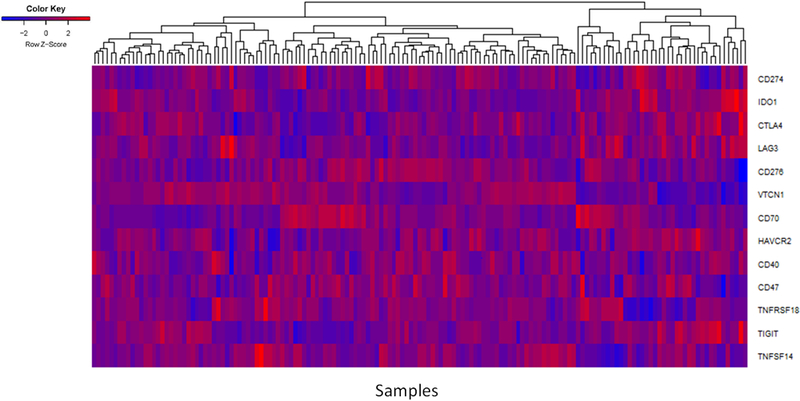
Immune-checkpoint therapy has failed to demonstrate meaningful clinical benefit in unselected cases of pancreatic adenocarcinoma (PDAC), but a subset of PDACs are known to upregulate pathways involved in acquired immune suppression. Further delineation of immunologic subtypes of PDAC is necessary to improve clinical trial designs and identify patients who might benefit from immune-checkpoint therapy. We used clinical survival and RNA expression data from The Cancer Genome Atlas (TCGA) to investigate the relationship between immune-modulating pathways and immune subset markers and their impact on survival in PDAC patients. Of the adaptive immune-resistance pathways, expression of PD-L1 and IDO1 was individually associated with poor survival. Although CD8 expression alone was not correlated with survival, the combination of PD-L1- and high CD8 expression identified a subtype with favorable survival. We further extended these observations using an independent PDAC cohort from our institution via IHC, again observing that the PD-L1-/CD8high subtype was associated with positive prognosis. Although PDAC is regarded as a poorly immunogenic cancer type, these findings infer that T-cell infiltration in the absence of adaptive immune-resistance pathways is a feature of long-term survival in PDAC and imply the importance of developing future immunotherapeutic strategies based on data-supported biomarkers to refine patient selection.
©2019 American Association for Cancer Research.
Conflict of interest statement
Figures


Similar articles
-
Programmed cell death ligand 1 cut-point is associated with reduced disease specific survival in resected pancreatic ductal adenocarcinoma.BMC Cancer. 2017 Sep 5;17(1):618. doi: 10.1186/s12885-017-3634-5. BMC Cancer. 2017. PMID: 28870260 Free PMC article.
-
PD-L1 expression in pancreatic ductal adenocarcinoma is a poor prognostic factor in patients with high CD8+ tumor-infiltrating lymphocytes: highly sensitive detection using phosphor-integrated dot staining.Int J Clin Oncol. 2017 Aug;22(4):726-733. doi: 10.1007/s10147-017-1112-3. Epub 2017 Mar 18. Int J Clin Oncol. 2017. PMID: 28314962 Free PMC article.
-
Analysis of PD-L1 promoter methylation combined with immunogenic context in pancreatic ductal adenocarcinoma.Cancer Immunol Immunother. 2024 Jun 4;73(8):149. doi: 10.1007/s00262-024-03745-y. Cancer Immunol Immunother. 2024. PMID: 38833018 Free PMC article.
-
The clinicopathological and prognostic significance of PD-L1 expression in pancreatic cancer: A meta-analysis.Hepatobiliary Pancreat Dis Int. 2018 Apr;17(2):95-100. doi: 10.1016/j.hbpd.2018.03.007. Epub 2018 Mar 13. Hepatobiliary Pancreat Dis Int. 2018. PMID: 29576277 Review.
-
Immune cell infiltrates as prognostic biomarkers in pancreatic ductal adenocarcinoma: a systematic review and meta-analysis.J Pathol Clin Res. 2021 Mar;7(2):99-112. doi: 10.1002/cjp2.192. Epub 2021 Jan 22. J Pathol Clin Res. 2021. PMID: 33481339 Free PMC article.
Cited by
-
Identification of the molecular subtype and prognostic characteristics of pancreatic cancer based on CD8 + T cell-related genes.Cancer Immunol Immunother. 2023 Mar;72(3):647-664. doi: 10.1007/s00262-022-03269-3. Epub 2022 Aug 29. Cancer Immunol Immunother. 2023. PMID: 36036290 Free PMC article.
-
Early Stage Finding of an Immune-Enhanced Genetic Subtype of Nonsmall Cell Lung Cancer Related with T-Cell Depletion.Evid Based Complement Alternat Med. 2022 Oct 14;2022:6765997. doi: 10.1155/2022/6765997. eCollection 2022. Evid Based Complement Alternat Med. 2022. PMID: 36276870 Free PMC article.
-
Single-cell dissection reveals the role of aggrephagy patterns in tumor microenvironment components aiding predicting prognosis and immunotherapy on lung adenocarcinoma.Aging (Albany NY). 2023 Dec 13;15(23):14333-14371. doi: 10.18632/aging.205306. Epub 2023 Dec 13. Aging (Albany NY). 2023. PMID: 38095634 Free PMC article.
-
Development of Prognostic Features of Hepatocellular Carcinoma Based on Metabolic Gene Classification and Immune and Oxidative Stress Characteristic Analysis.Oxid Med Cell Longev. 2023 Feb 18;2023:1847700. doi: 10.1155/2023/1847700. eCollection 2023. Oxid Med Cell Longev. 2023. PMID: 36860731 Free PMC article.
-
Integrated analysis of NET-DNA receptor CCDC25 in malignant tumors: Pan-cancer analysis.Health Sci Rep. 2023 Oct 12;6(10):e1621. doi: 10.1002/hsr2.1621. eCollection 2023 Oct. Health Sci Rep. 2023. PMID: 37841944 Free PMC article.
References
-
- Miller KD, Siegel RL, Lin CC, Mariotto AB, Kramer JL, Rowland JH, et al. Cancer treatment and survivorship statistics, 2016. CA Cancer J Clin. 2016;66:271–89. - PubMed
-
- Siegel RL, Miller KD, Jemal A. Cancer statistics, 2016. CA Cancer J Clin. 2016;66:7–30. - PubMed
-
- O’Reilly EM, Oh D-Y, Dhani N, Renouf DJ, Lee MA, Sun W, et al. A randomized phase 2 study of durvalumab monotherapy and in combination with tremelimumab in patients with metastatic pancreatic ductal adenocarcinoma (mPDAC): ALPS study J Clin Oncol. American Society of Clinical Oncology; 2018;36:217. - PubMed
MeSH terms
Substances
Grants and funding
LinkOut - more resources
Full Text Sources
Medical
Research Materials
