. 2019 May 1;79(9):2327-2338.
doi: 10.1158/0008-5472.CAN-18-1785.

Selective EGLN Inhibition Enables Ablative Radiotherapy and Improves Survival in Unresectable Pancreatic Cancer

Affiliations

Selective EGLN Inhibition Enables Ablative Radiotherapy and Improves Survival in Unresectable Pancreatic Cancer

Tara N Fujimoto et al. Cancer Res. .

Abstract

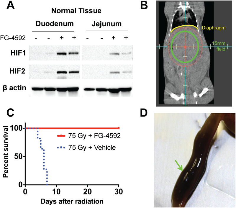
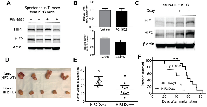
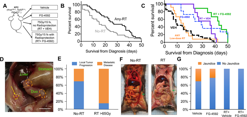
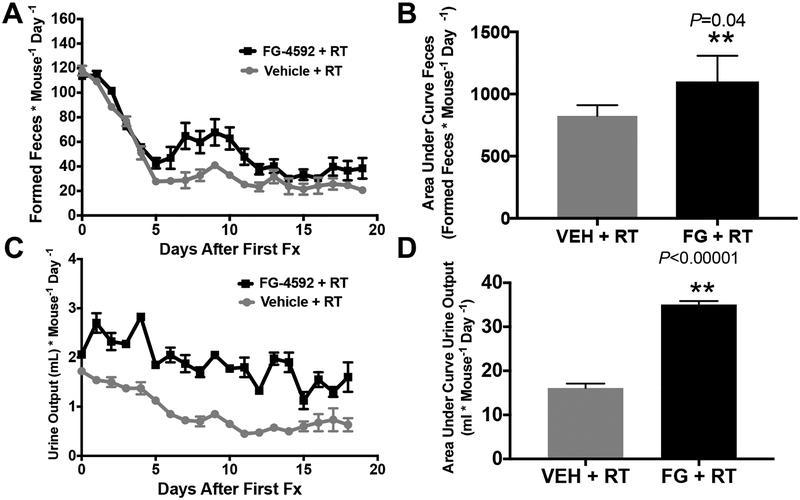
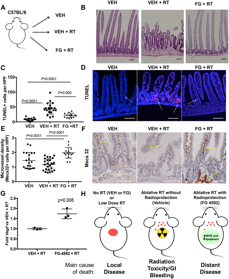
When pancreatic cancer cannot be removed surgically, patients frequently experience morbidity and death from progression of their primary tumor. Radiation therapy (RT) cannot yet substitute for an operation because radiation causes fatal bleeding and ulceration of the nearby stomach and intestines before achieving tumor control. There are no FDA-approved medications that prevent or reduce radiation-induced gastrointestinal injury. Here, we overcome this fundamental problem of anatomy and biology with the use of the oral EGLN inhibitor FG-4592, which selectively protects the intestinal tract from radiation toxicity without protecting tumors. A total of 70 KPC mice with autochthonous pancreatic tumors received oral FG-4592 or vehicle control ± ablative RT to a cumulative 75 Gy administered in 15 daily fractions to a limited tumor field. Although ablative RT reduced complications from local tumor progression, fatal gastrointestinal bleeding was observed in 56% of mice that received high-dose RT with vehicle control. However, radiation-induced bleeding was completely ameliorated in mice that received high-dose RT with FG-4592 (0% bleeding, P < 0.0001 compared with vehicle). Furthermore, FG-4592 reduced epithelial apoptosis by half (P = 0.002) and increased intestinal microvessel density by 80% compared with vehicle controls. EGLN inhibition did not stimulate cancer growth, as treatment with FG-4592 alone, or overexpression of HIF2 within KPC tumors independently improved survival. Thus, we provide a proof of concept for the selective protection of the intestinal tract by the EGLN inhibition to enable ablative doses of cytotoxic therapy in unresectable pancreatic cancer by reducing untoward morbidity and death from radiation-induced gastrointestinal bleeding. SIGNIFICANCE: Selective protection of the intestinal tract by EGLN inhibition enables potentially definitive doses of radiation therapy. This might allow radiation to be a surgical surrogate for unresectable pancreatic cancer.Graphical Abstract: http://cancerres.aacrjournals.org/content/canres/79/9/2327/F1.large.jpg.


Conflict of interest statement

Disclosures: The authors declare no conflicts of interest in this manuscript.

Figures

Figure 1.
Figure 1.. FG-4592 reduces morbidity from high-dose fractionated radiation
(A) Western blots demonstrating stabilized HIF in the duodenum and jejunum 6 hours after receiving FG-4592, representative of 4 mice per treatment condition. (B) Coronal image from a cone-beam CT taken during treatment with the circular 15mm field superimposed. Diaphragm (yellow) is pointed out for reference. (C) Improved survival of mice treated with FG-4592 after 75Gy/15fx of UA-XRT (n=8–10/tx cohort, Log rank p<0.0001). (D) Example of a duodenum lumen filled with partially digested blood (green arrow) as a result of radiation toxicity.
Figure 2.
Figure 2.. HIF stabilization within KPC tumors does not enhance tumor growth.
(A) EGLN inhibition by FG-4592 does not increase HIF expression by Western Blot, representative of 4–6 tumors per treatment group, with quantified relative levels of HIF1 and HIF2 in (B, error bars represent s.e.m.). (C) Overexpression of HIF2 in syngeneic orthotopic KPC tumors confirmed by Western Blot which are representative of 4 tumors from each treatment group. HIF2 expression decreased tumor growth as determined by visual inspection (D, scale bar=10mm) and tumor weight (E, n=9–10/cohort; unpaired two-tailed t-test *P=0.0004, error bars are s.e.m.), contributing to improved survival (F, n=11 per cohort, log rank P<0.0001)
Figure 3.
Figure 3.. High-dose radiation with concurrent FG-4592 improves survival and reduces clinical signs of local progression
(A) Experimental scheme for KPC animals. (B) Kaplan-Meier survival curves for mice who received any radiation (any-RT= RT+VEH or RT+FG) or no radiation therapy (No-RT=VEH or FG) as part of their therapy. Median survival was higher in Any-RT group compared to No-RT (Gehan-Breslow-Wilcoxan P=0.006). (C) Kaplan-Meier survival curves for all animals, with highest overall survival for mice treated with RT + FG-4592 (Log rank P<.0001). (D) Gross photograph of the abdominal cavity of a mouse with local tumor dissemination. The wall of the gastric antrum (orange dashed line) and duodenum is thickened by invading cancer cells, which have obstructed gastric outflow leading to gastric bloating. There is a single tumor nodule (yellow arrow) in the peritoneal wall. Scale bar=5mm (E) Local tumor progression more commonly necessitated euthanasia in No-RT treatment groups compared to those that received >65Gy radiation treatment (Fisher’s exact P<0.001) (F) An illustrative example of jaundice. Icteric KPC mouse that had no RT (left image) and an non-icteric KPC mouse treated with RT (right image). The pancreas (black arrow), subcutaneous fat (green arrow) and the gallbladder (blue arrow) are shown in both mice. Scale bar=1cm (G) Mice who did not receive RT had higher rates of jaundice (Chi-square P=0.02)
Figure 4.
Figure 4.. EGLN inhibition by FG-4592 reduces toxicity and enables definitive radiation treatment of pancreatic cancer.
Distribution of all causes of morbidity leading to death for (A) Vehicle alone, (B) FG-4592 alone, (C) >65Gy RT + Vehicle and (D) >65Gy RT + FG-4592. (E) An example of intestinal bleeding (arrows) observed at necropsy in a mouse that received 75Gy of fractionated radiation without radioprotection (scale bar=1cm) with (F) histopathologic correlation from the same mouse showing edema, hemorrhage, and leukocytic infiltration. (G) Representative histopathology of a jejunal section at necropsy from mouse treated with full dose ablative RT with FG-4592 for radioprotection. The major lesions identified were edema, focal crypt necrosis, and leukocytic infiltration.
Figure 5.
Figure 5.. Metabolic cage analysis of mice treated with a clinically relevant course of dose-escalated RT to a limited upper abdominal field.
(A) Formed feces during fractionated radiation treatments with area under the curve quantification in (B). More formed feces denotes less diarrhea, as loose stools cannot be quantified in metabolic cage method. (C) Urine output during fractionated radiation treatments with area under the curve quantification in (D).
Figure 6.
Figure 6.. Mechanisms of FG-4592 Intestinal radioprotection.
(A) Scheme of experimental groups, where mice received daily vehicle alone (VEH), vehicle with 75Gy/15 fractions of radiation (VEH +RT) or FG-4592 with the same radiation treatment (FG +RT). Intestines from the proximal duodenum to the second portion of the jejunum were collected and analyzed over serial ongitudinal sections (see methods). (B) Representative images of lesions observed from H&E-stained small intestines of VEH (n=5), 75Gy/15 fractions (n=5), and 75Gy/15 fraction + FG-4592 treated mice (n=5) observed in the duodenum of RT+VEH group (2 of 5 animals), but not in other cohorts. These lesions are characterized by villi blunting and crypt hypertrophy. Scale bars=100 μm (C) TUNEL+ cells per HPF in the small intestines of the indicated groups with representative examples in (D). Scale bars=50 μm (E) Quantification of Meca-32 staining for endothelial cells in the epithelia of the small intestines within the three cohorts, quantified on 3–5 HPF per mouse, with representative images used for quantification in (F). Scale bars=50 μm. (G) Jejunal epithelium was isolated from each of the cohorts and assessed by quantitative PCR for relative mRNA levels of Vegf. (H) Schematic of rationale for radiation protection with high-dose radiation for unresectable pancreatic cancer. Radiation slows local progression, but often causes toxicity, but a combination of high-dose radiation along with FG-4592 for radioprotection provides local control while preventing toxicity from treatment, contributing to survival.

References

    1. Rahib L, Smith BD, Aizenberg R, Rosenzweig AB, Fleshman JM, Matrisian LM. Projecting cancer incidence and deaths to 2030: the unexpected burden of thyroid, liver, and pancreas cancers in the United States. Cancer Res 2014;74:2913–21 - PubMed
    1. Balaban EP, Mangu PB, Khorana AA, Shah MA, Mukherjee S, Crane CH, et al. Locally Advanced, Unresectable Pancreatic Cancer: American Society of Clinical Oncology Clinical Practice Guideline. J Clin Oncol 2016;34:2654–68 - PubMed
    1. Iacobuzio-Donahue CA, Fu B, Yachida S, Luo M, Abe H, Henderson CM, et al. DPC4 gene status of the primary carcinoma correlates with patterns of failure in patients with pancreatic cancer. J Clin Oncol 2009;27:1806–13 - PMC - PubMed
    1. Kelly P, Das P, Pinnix CC, Beddar S, Briere T, Pham M, et al. Duodenal toxicity after fractionated chemoradiation for unresectable pancreatic cancer. International journal of radiation oncology, biology, physics 2013;85:e143–9 - PubMed
    1. Krishnan S, Chadha AS, Suh Y, Chen HC, Rao A, Das P, et al. Focal Radiation Therapy Dose Escalation Improves Overall Survival in Locally Advanced Pancreatic Cancer Patients Receiving Induction Chemotherapy and Consolidative Chemoradiation. Int J Radiat Oncol Biol Phys 2016;94:755–65 - PMC - PubMed

Publication types

MeSH terms