. 2019 May 1;11(490):eaat8329.
doi: 10.1126/scitranslmed.aat8329.

Quorum sensing between bacterial species on the skin protects against epidermal injury in atopic dermatitis

Affiliations

Quorum sensing between bacterial species on the skin protects against epidermal injury in atopic dermatitis

Michael R Williams et al. Sci Transl Med. .

Abstract

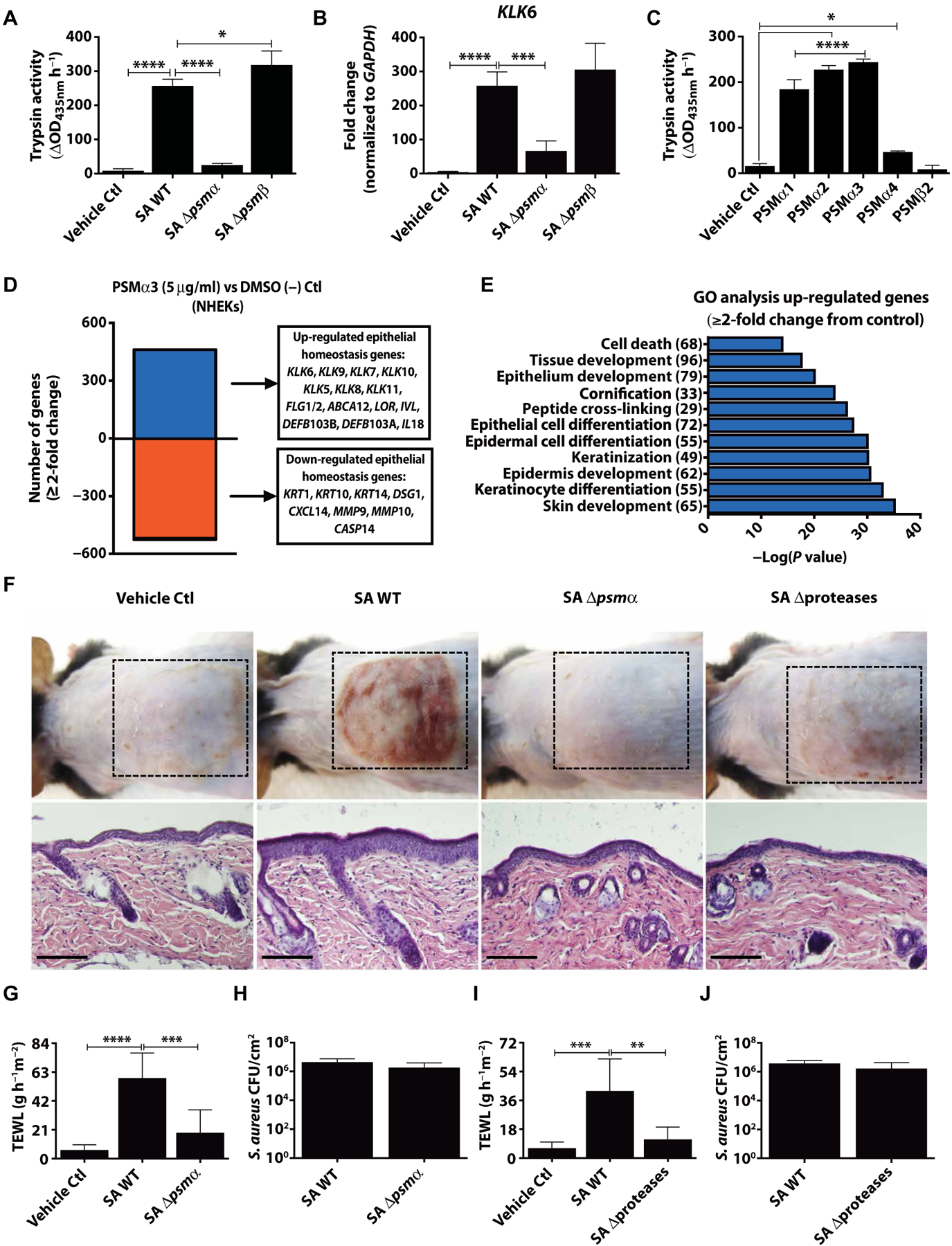
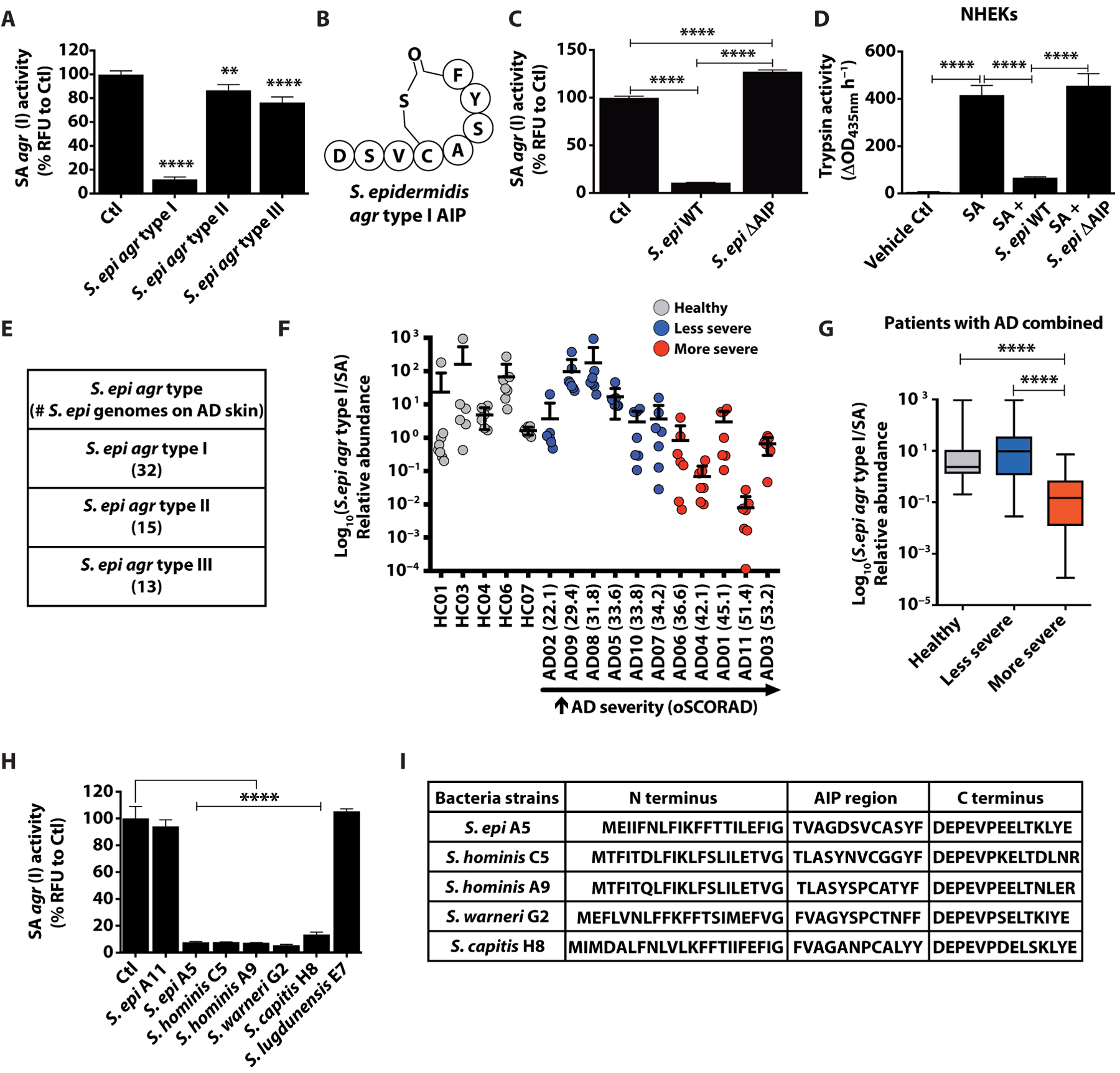
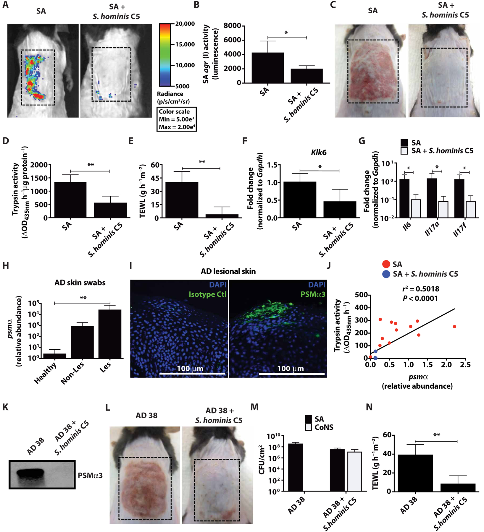
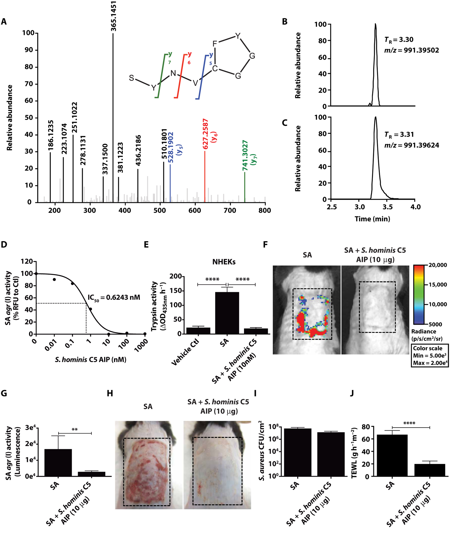
Colonization of the skin by Staphylococcus aureus is associated with exacerbation of atopic dermatitis (AD), but any direct mechanism through which dysbiosis of the skin microbiome may influence the development of AD is unknown. Here, we show that proteases and phenol-soluble modulin α (PSMα) secreted by S. aureus lead to endogenous epidermal proteolysis and skin barrier damage that promoted inflammation in mice. We further show that clinical isolates of different coagulase-negative staphylococci (CoNS) species residing on normal skin produced autoinducing peptides that inhibited the S. aureus agr system, in turn decreasing PSMα expression. These autoinducing peptides from skin microbiome CoNS species potently suppressed PSMα expression in S. aureus isolates from subjects with AD without inhibiting S. aureus growth. Metagenomic analysis of the AD skin microbiome revealed that the increase in the relative abundance of S. aureus in patients with active AD correlated with a lower CoNS autoinducing peptides to S. aureus ratio, thus overcoming the peptides' capacity to inhibit the S. aureus agr system. Characterization of a S. hominis clinical isolate identified an autoinducing peptide (SYNVCGGYF) as a highly potent inhibitor of S. aureus agr activity, capable of preventing S. aureus-mediated epithelial damage and inflammation on murine skin. Together, these findings show how members of the normal human skin microbiome can contribute to epithelial barrier homeostasis by using quorum sensing to inhibit S. aureus toxin production.


Conflict of interest statement

Competing interests: M.R.W. and R.L.G. are co-inventors of UCSD technology related to the bacterial quorum sensing inhibition therapy discussed here under patent PCT/US18/49237 titled “Molecular bacteriotherapy to control skin enzymatic activity.” R.L.G. is a co-founder, scientific advisor, consultant, and has equity in MatriSys Biosciences, and is a consultant, receives income from, and has equity in Sente.

Figures

Fig. 1.
Fig. 1.. S. aureus PSMα induces keratinocyte protease activity and disrupts epithelial barrier homeostasis.
NHEKs were stimulated with 5% S. aureus USA300 LAC (SA) supernatant from overnight cultures [1 × 10⁹ colony-performing units (CFU)] of wild-type (WT) and psmα (Δpsmα) or psmβ (Δpsmβ) knockout strains for 24 hours. Both (A) trypsin activity and (B) KLK6 mRNA expression compared to the housekeeping gene GAPDH were analyzed (n = 4). (C) PSM synthetic peptides were added to NHEKs for 24 hours to analyze changes in trypsin activity (n = 4). (D) Transcript analysis by RNA-seq and (E) gene ontology (GO) analysis of genes that changed ≥2-fold 24 hours after PSMα3 treatment. The number of genes in each category shown in parentheses. (F) Eight-week-old male C57BL/6 mice were treated for 72 hours with 1 × 10⁷ CFU SA WT (USA300 LAC) and corresponding SA Δpsmα, or SA USA300 LAC WT (AH1263) and corresponding SA Δproteases [SA WT in (F) representative of both SA WT strains]. Bacterial growth medium was used as a vehicle control. Dashed lines indicate treatment area. Scale bars, 200 μm (n = 6). (G to J) Transepidermal water loss (TEWL) or measurement of CFU/cm2 on mice after 72-hour treatment with initial 1 × 10⁷ CFU of (G and H) SA WT (USA300 LAC) and corresponding SA Δpsmα or (I and J) SA WT [USA300 LAC (AH1263)] and corresponding SA Δproteases (n = 6). All experiments are representative of three independent experiments. Error bars are SEM. One-way ANOVA was used to determine statistical significance. *P < 0.05, **P < 0.01, ***P < 0.001, ****P < 0.0001. Ctl, control.
Fig. 2.
Fig. 2.. S. epidermidis agr type I autoinducing peptide characterization and deficiency in AD skin.
(A) SA USA300 LAC agr type I P3-YFP activity after overnight culture (1 × 10⁹ CFU) with 25% supernatant from overnight culture of S. epidermidis (S. epi) agr types I (RP62A), II (1457), and III (8247) (n = 4). (B) Structure of S. epidermidis agr type I autoinducing peptide (AIP). (C) Analysis of agr activity as in (A) with S. epidermidis agr type I (RP62A) WT or autoinducing peptide knockout (ΔAIP) strain supernatants (n = 4). (D) NHEK trypsin activity after culture for 24 hours with 5% SA supernatant grown overnight with or without S. epidermidis agr type I (RP62A) WT or autoinducing peptide knockout (ΔAIP) (n = 4). (E) Number (#) of S. epidermidis agr types I to III strains found on AD skin from metagenomic dataset. (F and G) Ratio of S. epidermidis agr type I to SA relative abundance on the combined analysis of all sites sampled from 5 healthy individuals and 11 AD subjects during flare and ranked from “least severe” to “most severe” based on oSCORAD. (H) Analysis of agr activity as in (A) after coculture overnight with 25% supernatant of overnight cultures (1 × 10⁹ CFU) of clinical CoNS strains (n = 3). (I) Assessment of autoinducing peptide coding regions in agrD gene of the inhibitory CoNS strains. All experiments are representative of three independent experiments. All error bars are SEM. One-way ANOVA or an (nonparametric) unpaired Kruskal-Wallis test in (G) was used to determine statistical significance. *P < 0.05, **P < 0.01, ***P < 0.001, ****P < 0.0001.
Fig. 3.
Fig. 3.. Inhibition of S. aureus agr activity by clinical S. hominis isolate correlates with prevention of skin barrier damage and inflammation.
(A to C) SA USA300 LAC agr type I pAmi P3-Lux reporter strain (AH2759) (1 × 10⁷ CFU) combined with or without live S. hominis C5 (1 × 10⁷ CFU) was applied to murine back skin for 72 hours, and SA agr activity was assessed by changes in luminescence. Images are representative of n = 5; dashed boxes indicate treatment area. (D to G) Murine back skin after 72-hour bacteria treatment, assessed for changes in TEWL, trypsin activity, and Klk6 and cytokine (Il6, Il17a, and Il17f) mRNA expression normalized to housekeeping gene Gapdh (n = 5). (H) Relative abundance of SA psmα mRNA isolated from swabs of healthy (n = 6) and AD nonlesional and lesional skin (n = 5). (I) Detection of SA PSMα3 by immunofluorescence in epidermis of AD lesional skin (representative image with scale bar, 100 μm). (J) Clinical SA isolates from 11 patients were grown up to 18 hours (1 × 10⁹ CFU) with or without 25% S. hominis C5 supernatant. psmα mRNA expression was measured at 8 hours, whereas trypsin activity was assessed from NHEKs treated 24 hours with 18-hour cultured SA (5%) supernatant (n = 3). (K) Immunoblot of culture supernatant from clinical SA isolate AD 38 grown overnight (1 × 10⁹ CFU) with or without S. hominis C5 supernatant and probed for PSMα3. (L to N) SA AD 38 (1 × 10⁷ CFU) was applied to murine back skin with or without S. hominis C5 (1 × 10⁷ CFU) for 72 hours (n = 5) (dashed boxes indicate treatment area). Changes in CFU/cm2 of SA and CoNS, and TEWL are shown. All experiments are representative of two independent experiments. Error bars represent SEM. Student’s t tests, Pearson correlation coefficient (J), and an (nonparametric) unpaired Kruskal-Wallis test (H) were used to determine statistical significance. *P < 0.05, **P < 0.01, ***P < 0.001, ****P < 0.0001.
Fig. 4.
Fig. 4.. Identification of an autoinducing peptide from a clinical S. hominis isolate that inhibits S. aureus agr activity.
(A) Determination of S. hominis C5 autoinducing peptide sequence as SYNVCGGYF. The MS-MS spectrum obtained with the synthetic peptide is shown. Bolded fragment peaks and those indicated in color were identified in the MS-MS spectra for both the synthetic peptide and the spent growth medium sample (with difference in mass of <5 parts per million). Only matching fragments above 15% abundance are shown. Key features used for identification were the y5 (blue), y6 (red), and y7 (green) fragments of the cyclic autoinducing peptide. The selected-ion chromatograms for mass/charge ratio (m/z) 991.3984 ± 5 ppm for (B) bacteria culture and (C) synthetic autoinducing peptide in growth medium confirmed the retention time (TR) and accurate m/z of the predicted autoinducing peptide sequence. (D) Analysis of S. hominis C5 synthetic autoinducing peptide on SA USA300 LAC agr type I P3-YFP activity after an overnight culture (1 × 10⁹ CFU) and IC50 value indicated by dotted lines at midpoint of the curve. (E) Measurement of NHEK trypsin activity after 24-hour incubation with 5% supernatant from overnight culture of SA with or without S. hominis C5 autoinducing peptide (1 × 10⁹ CFU) (n = 4). (F to H) SA USA300 LAC agr type I pAmi P3-Lux with or without S. hominis C5 synthetic autoinducing peptide was colonized (1 × 10⁷ CFU) on murine back skin for 48 hours followed by analysis of agr activity (luminescence) and representative pictures of murine back skin after treatment (dashed lines represent treated areas) (n = 5). (I and J) SA CFU/cm2 and TEWL after treatment with S. hominis C5 synthetic autoinducing peptide. Data are representative of two experiments. Error bars are SEM. One-way ANOVA and Student’s t tests were used to determine statistical significance. *P < 0.05, **P < 0.01, ***P < 0.001, ****P < 0.0001.

References

    1. Eckert L, Gupta S, Amand C, Gadkari A, Mahajan P, Gelfand JM, The burden of atopic dermatitis in US adults: Health care resource utilization data from the 2013 National Health and Wellness Survey. J. Am. Acad. Dermatol 78, 54–61.e1 (2018). - PubMed
    1. Narla S, Hsu DY, Thyssen JP, Silverberg JI, Inpatient financial burden of atopic dermatitis in the United States. J. Invest. Dermatol 137, 1461–1467 (2017). - PubMed
    1. Palmer CN, Irvine AD, Terron-Kwiatkowski A, Zhao Y, Liao H, Lee SP, Goudie DR, Sandilands A, Campbell LE, Smith FJ, O’Regan GM, Watson RM, Cecil JE, Bale SJ, Compton JG, DiGiovanna JJ, Fleckman P, Lewis-Jones S, Arseculeratne G, Sergeant A, Munro CS, El Houate B, McElreavey K, Halkjaer LB, Bisgaard H, Mukhopadhyay S, McLean WH, Common loss-of-function variants of the epidermal barrier protein filaggrin are a major predisposing factor for atopic dermatitis. Nat. Genet 38, 441–446 (2006). - PubMed
    1. De Benedetto A, Rafaels NM, McGirt LY, Ivanov AI, Georas SN, Cheadle C, Berger AE, Zhang K, Vidyasagar S, Yoshida T, Boguniewicz M, Hata T, Schneider LC, Hanifin JM, Gallo RL, Novak N, Weidinger S, Beaty TH, Leung DY, Barnes KC, Beck LA, Tight junction defects in patients with atopic dermatitis. J. Allergy Clin. Immunol 127, 773–786.e1-7 (2011). - PMC - PubMed
    1. Kato A, Fukai K, Oiso N, Hosomi N, Murakami T, Ishii M, Association of SPINK5 gene polymorphisms with atopic dermatitis in the Japanese population. British J. Dermatol 148, 665–669 (2003). - PubMed

Publication types