. 2019 May 1;9(1):6769.
doi: 10.1038/s41598-019-43293-5.

Prognostic role of ARID1A negative expression in gastric cancer

Affiliations

Prognostic role of ARID1A negative expression in gastric cancer

Mai Ashizawa et al. Sci Rep. .

Abstract

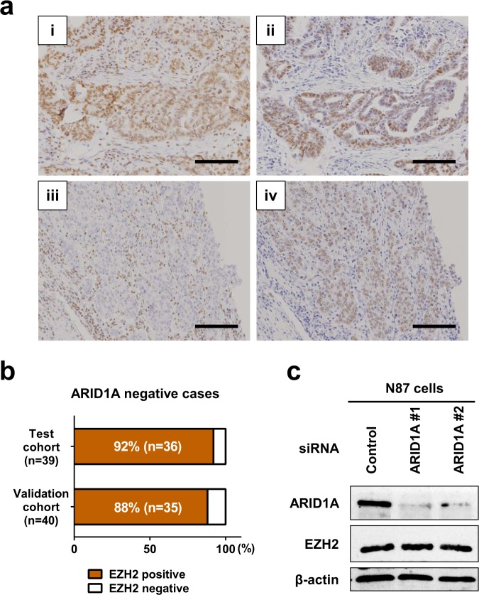
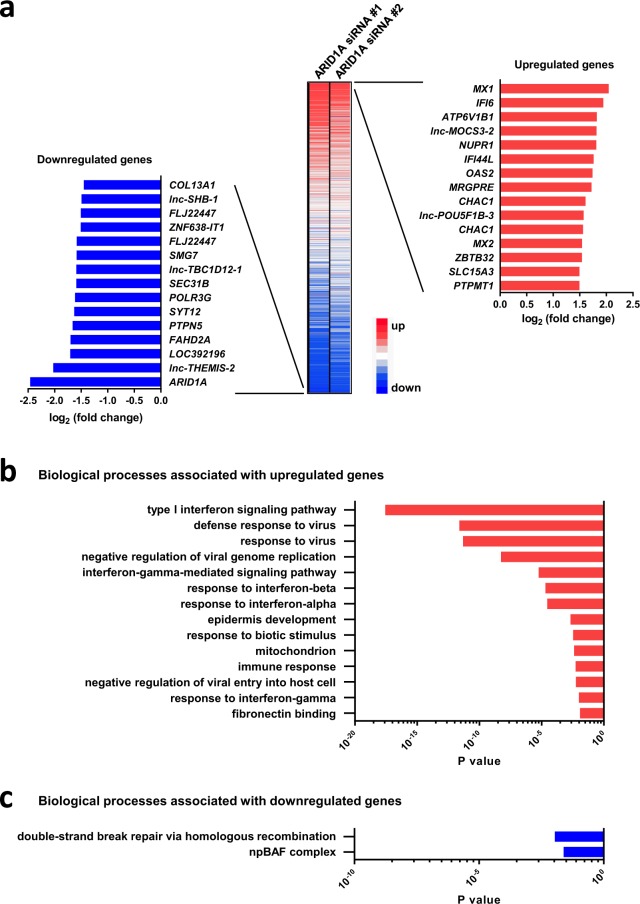
AT-rich interactive domain 1A (ARID1A) functions as a tumor suppressor and several therapeutic targets in ARID1A-mutated cancers are under development. Here, we investigated the prognostic value of ARID1A for gastric cancer and its association with expression of PD-L1 and p53. ARID1A expression was examined by immunohistochemistry and negative expression of ARID1A was detected in 39 (19.5%) of 200 cases in a test cohort and in 40 (18.2%) of 220 cases in a validation cohort. Negative expression of ARID1A was associated with worse overall survival in undifferentiated cases, particularly early-stage cases. Negative expression of ARID1A was detected in 11 (50%) of 22 PD-L1-positive cases and in 68 (17.1%) of 398 PD-L1-negative cases in a combined cohort. Negative expression of ARID1A was detected in 45 (22%) of 205 p53-positive cases and in 34 (15.8%) of 215 p53-negative cases in a combined cohort. In addition, expression of EZH2, a potential synthetic lethal target in ARID1A-mutated tumors, was detected in 79 ARID1A-negative cases. An ARID1A-knockdown gastric cancer cell line was subjected to microarray analysis, but no actionable targets or pathways were identified. The present results indicate that ARID1A may serve as an early-stage prognostic biomarker for undifferentiated gastric cancer.


Conflict of interest statement

The authors declare no competing interests.

Figures

Figure 1
Figure 1
Representative images showing immunohistochemical staining for ARID1A in gastric cancer (a–h). (a,c) Positive ARID1A staining in differentiated tumor tissues. (b,d) Negative ARID1A staining in differentiated tumor tissues. (e,g) Positive ARID1A staining in undifferentiated tumor tissues. (f,h) Negative ARID1A staining in undifferentiated tumor tissues. Scale bars = 100 μm.
Figure 2
Figure 2
Kaplan-Meier curves showing overall survival of patients with gastric cancer according to ARID1A expression. Kaplan-Meier survival analysis of all cases, differentiated cases, and undifferentiated cases in the test (a), validation (b), and combined cohorts (c).
Figure 3
Figure 3
Kaplan-Meier curves showing overall survival of patients with gastric cancer according to ARID1A expression. (a) Kaplan-Meier curves showing overall survival of the patients with gastric cancer according to ARID1A expression. Kaplan-Meier survival analysis of all cases in the combined cohort (n = 420) and of TNM stage 1 (n = 212), stage 2 (n = 79), stage 1&2 (n = 291), and stage 3&4 (n = 129) cases stratified according to ARID1A expression. (b) Kaplan-Meier curves showing overall survival of patients with gastric cancer according to ARID1A expression. Kaplan-Meier survival analysis of undifferentiated cases in the combined cohorts (n = 205) and of TNM stage 1 (n = 88), stage 2 (n = 41), stage 1&2 (n = 129), and stage 3&4 (n = 76) cases stratified according to ARID1A expression.
Figure 4
Figure 4
Association between expression of ARID1A and PD-L1 and p53 in gastric cancer. (a) Representative images showing immunohistochemical staining of gastric cancer tissue for PD-L1 (i–vi). (i, ii) A case showing positive ARID1A (i) and positive PD-L1 (ii) staining in differentiated tumor tissues. (iii, iv) A case showing positive ARID1A (iii) and positive PD-L1 (iv) staining in undifferentiated tumor tissues. (v, vi) A case showing negative ARID1A (v) and positive PD-L1 (vi) staining in undifferentiated tumor tissues. Scale bars = 100 μm. (b) Differences in PD-L1 expression between ARID1A-positive and -negative gastric cancer in the combined cohorts. P = 0.0007, Fisher’s exact test. (c) PD-L1 mRNA expression in gastric cancer cell lines with ARID1A truncating mutations (Mut, n = 6) and the wild-type (WT, n = 28). The average expression level of PD-L1 was higher in ARID1A Mut cell lines than in ARID1A WT cell lines. Data were obtained from the Cancer Cell Line Encyclopedia (CCLE). P = 0.297, Mann Whitney test. (d) PD-L1 mRNA expression in gastric cancer with ARID1A truncating mutations (Mut, n = 83) and wild-type ARID1A (WT, n = 286). The average expression level of PD-L1 was higher in ARID1A Mut than in ARID1A WT cases. Data were provided by The Cancer Genome Atlas (TCGA [Provisional]). P < 0.0001, Mann Whitney test. (e) Representative immunohistochemical staining for p53 in gastric cancer. Positive and negative p53 staining in tumor tissues. (i, ii) A case showing positive ARID1A (i) and positive p53 (ii) staining in differentiated tumor tissues. (iii, iv) A case showing positive ARID1A (iii) and negative p53 (iv) staining in undifferentiated tumor tissues. Scale bars = 100 μm. (f) Differences in p53 expression between ARID1A-positive and -negative gastric cancer in the combined cohorts. Not significant, Fisher’s exact test. (g) Comparison of ARID1A mutations and TP53 mutations in gastric cancer in the TCGA cohort. TP53 was less frequently mutated in gastric cancer with ARID1A truncating mutations. P = 0.021, Fisher’s exact test.
Figure 5
Figure 5
Association between EZH2 and ARID1A expression in gastric cancer. (a) Representative images showing immunohistochemical staining of EZH2 in gastric cancer. (i, ii) A case showing positive ARID1A (i) and positive EZH2 (ii) staining in differentiated tumor tissues. (iii, iv) A case showing negative ARID1A (iii) and positive EZH2 (iv) staining in undifferentiated tumor tissues. Scale bars = 100 μm. (b) EZH2 expression in ARID1A-negative gastric cancer in the test (n = 39) and validation (n = 40) cohorts. (c) Western blot analysis of ARID1A and EZH2 expression in N87 cells in which ARID1A was silenced using two different siRNAs (#1 and #2). ARID1A was downregulated, whereas EZH2 was positively detected. β-actin was used as a loading control. The same cells were used for the microarray analysis in Fig. 6a.
Figure 6
Figure 6
Functional annotation analysis using genes altered by ARID1A knockdown in N87 cells. (a) Heatmap showing gene alterations in two different ARID1A knockdown cells relative to control (scrambled siRNA) cells. Two different siRNAs (#1 and #2) were used. The 15 genes showing the greatest upregulation and downregulation are highlighted. (b) Gene ontology (GO) terms (biological processes) significantly enriched among upregulated genes in ARID1A knockdown cells relative to control (scrambled siRNA) cells, as determined by DAVID functional annotation analysis. (c) GO terms significantly enriched among downregulated genes in ARID1A knockdown cells relative to control (scrambled siRNA) cells, as determined by DAVID functional annotation analysis.

References

    1. Mathur R, et al. ARID1A loss impairs enhancer-mediated gene regulation and drives colon cancer in mice. Nat Genet. 2017;49:296–302. doi: 10.1038/ng.3744. - DOI - PMC - PubMed
    1. Zang ZJ, et al. Exome sequencing of gastric adenocarcinoma identifies recurrent somatic mutations in cell adhesion and chromatin remodeling genes. Nat Genet. 2012;44:570–574. doi: 10.1038/ng.2246. - DOI - PubMed
    1. Wang K, et al. Exome sequencing identifies frequent mutation of ARID1A in molecular subtypes of gastric cancer. Nat Genet. 2011;43:1219–1223. doi: 10.1038/ng.982. - DOI - PubMed
    1. Cancer Genome Atlas Research, N. Comprehensive molecular characterization of gastric adenocarcinoma. Nature513, 202−209:10.1038/nature13480 (2014). - PMC - PubMed
    1. Abe H, et al. ARID1A expression loss in gastric cancer: pathway-dependent roles with and without Epstein-Barr virus infection and microsatellite instability. Virchows Arch. 2012;461:367–377. doi: 10.1007/s00428-012-1303-2. - DOI - PubMed

Publication types

MeSH terms