Bilateral Syphilitic Optic Neuropathy with Secondary Autoimmune Optic Neuropathy and Poor Visual Outcome
- PMID: 31043959
- PMCID: PMC6477499
- DOI: 10.1159/000496142
Bilateral Syphilitic Optic Neuropathy with Secondary Autoimmune Optic Neuropathy and Poor Visual Outcome
Abstract
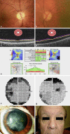
We describe the case of a 65-year-old man who suffered progressive visual loss despite appropriate treatment of ocular syphilis. Our patient initially presented with a unilateral 6th nerve palsy and associated double vision, which self-resolved over 6 months. His ophthalmic examination was otherwise normal. 12 months after the initial complaint, he represented with dyschromatopsia, reduced visual acuity, tonic pupils, and optic nerve atrophy. He tested positive for syphilis and was admitted for treatment of neurosyphilis with high-dose benzylpenicillin. Despite treatment, at a 4-month review his visual acuity remained poor and progression of optic nerve atrophy was noted alongside the development of bilateral central scotomas. Further testing was congruent with a diagnosis of autoimmune optic retinopathy. We propose this to be secondary to his syphilitic infection. Syphilis is known as the "great mimicker," and despite being quite treatable, this case highlights ongoing complexity in the diagnosis and management of syphilis, unfortunately with a poor visual outcome.
Keywords: Autoimmune optic neuropathy; Neurosyphilis; Optic neuropathy; Syphilis; Treponema pallidum.
Figures

Similar articles
-
Neurosyphilis presenting as visually asymptomatic bilateral optic perineuritis.BMJ Case Rep. 2019 Dec 22;12(12):e232520. doi: 10.1136/bcr-2019-232520. BMJ Case Rep. 2019. PMID: 31871011 Free PMC article.
-
Presentation of Ocular Syphilis with Bilateral Optic Neuropathy.Neuroophthalmology. 2023 Jul 5;47(5-6):274-280. doi: 10.1080/01658107.2023.2222800. eCollection 2023. Neuroophthalmology. 2023. PMID: 38130808 Free PMC article.
-
Isolated presumed optic nerve gumma, a rare presentation of neurosyphilis.Am J Ophthalmol Case Rep. 2017 Feb 3;6:7-10. doi: 10.1016/j.ajoc.2017.01.003. eCollection 2017 Jun. Am J Ophthalmol Case Rep. 2017. PMID: 29260044 Free PMC article.
-
Syphilis: reemergence of an old adversary.Ophthalmology. 2006 Nov;113(11):2074-9. doi: 10.1016/j.ophtha.2006.05.048. Epub 2006 Aug 28. Ophthalmology. 2006. PMID: 16935333 Review.
-
Emerging syphilitic optic neuropathy: critical review and recommendations.Restor Neurol Neurosci. 2008;26(4-5):279-89. Restor Neurol Neurosci. 2008. PMID: 18997306 Review.
Cited by
-
Neurosyphilis presenting as visually asymptomatic bilateral optic perineuritis.BMJ Case Rep. 2019 Dec 22;12(12):e232520. doi: 10.1136/bcr-2019-232520. BMJ Case Rep. 2019. PMID: 31871011 Free PMC article.
-
Presentation of Ocular Syphilis with Bilateral Optic Neuropathy.Neuroophthalmology. 2023 Jul 5;47(5-6):274-280. doi: 10.1080/01658107.2023.2222800. eCollection 2023. Neuroophthalmology. 2023. PMID: 38130808 Free PMC article.
-
Dyschromatopsia and contrast sensitivity changes in COVID-19 patients.Indian J Ophthalmol. 2024 May 1;72(5):664-671. doi: 10.4103/IJO.IJO_1437_23. Epub 2023 Dec 26. Indian J Ophthalmol. 2024. PMID: 38153970 Free PMC article.
References
-
- McLeish WM, Pulido JS, Holland S, Culbertson WW, Winward K. The ocular manifestations of syphilis in the human immunodeficiency virus type 1-infected host. Ophthalmology. 1990 Feb;97((2)):196–203. - PubMed
-
- Hausman L. The surgical treatment of syphilitic optic atrophy due to chiasmal arachnoiditis. Am J Ophthalmol. 1941;24((2)):119–32.
Publication types
LinkOut - more resources
Full Text Sources

