. 2019 Apr 25:7:12.
doi: 10.1038/s41413-019-0051-1. eCollection 2019.

DMP1 prevents osteocyte alterations, FGF23 elevation and left ventricular hypertrophy in mice with chronic kidney disease

Affiliations

DMP1 prevents osteocyte alterations, FGF23 elevation and left ventricular hypertrophy in mice with chronic kidney disease

Corey Dussold et al. Bone Res. .

Abstract

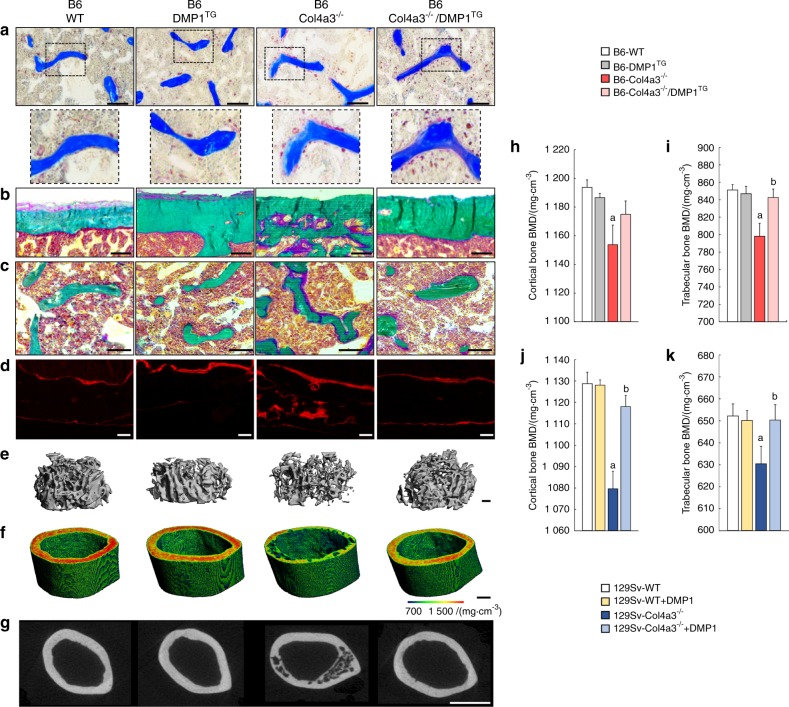
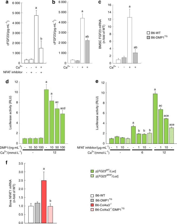
During chronic kidney disease (CKD), alterations in bone and mineral metabolism include increased production of the hormone fibroblast growth factor 23 (FGF23) that may contribute to cardiovascular mortality. The osteocyte protein dentin matrix protein 1 (DMP1) reduces FGF23 and enhances bone mineralization, but its effects in CKD are unknown. We tested the hypothesis that DMP1 supplementation in CKD would improve bone health, prevent FGF23 elevations and minimize consequent adverse cardiovascular outcomes. We investigated DMP1 regulation and effects in wild-type (WT) mice and the Col4a3-/- mouse model of CKD. Col4a3-/- mice demonstrated impaired kidney function, reduced bone DMP1 expression, reduced bone mass, altered osteocyte morphology and connectivity, increased osteocyte apoptosis, increased serum FGF23, hyperphosphatemia, left ventricular hypertrophy (LVH), and reduced survival. Genetic or pharmacological supplementation of DMP1 in Col4a3-/- mice prevented osteocyte apoptosis, preserved osteocyte networks, corrected bone mass, partially lowered FGF23 levels by attenuating NFAT-induced FGF23 transcription, and further increased serum phosphate. Despite impaired kidney function and worsened hyperphosphatemia, DMP1 prevented development of LVH and improved Col4a3-/- survival. Our data suggest that CKD reduces DMP1 expression, whereas its restoration represents a potential therapeutic approach to lower FGF23 and improve bone and cardiac health in CKD.

Keywords: Bone; Calcium and phosphate metabolic disorders; Pathogenesis.


Conflict of interest statement

V.D. has served as a consultant or received honoraria from Vifor, Luitpold, and grant support from Keryx Biopharmaceuticals and Vifor. M.W. has served as a consultant or received honoraria from Amag, Amgen, Akebia, Ardelyx, Diasorin, Keryx, Luitpold, and Sanofi, and grant support from Shire. The remaining authors have no competing interests.

Figures

Fig. 1
Fig. 1
DMP1 deficiency and supplementation in Col4a3−/− mice with advanced CKD. ab DMP1 mRNA expression in whole bone, kidney and heart of a 20 week-old B6 WT, DMP1TG, Col4a3−/−, and Col4a3−/−/DMP1TG mice and whole bone of b 9-week-old 129sv WT and Col4a3−/− mice treated with mouse recombinant his-tagged DMP1 or saline control once a day for one week. c Bright-field microscopy of DMP1 immunostaining in cortical bone of B6 WT and Col4a3−/− mice (scale bar = 50 µm). d His-tag specific immunodetection of DMP1 in bone of 129 Sv WT and Col4a3−/− mice injected with mouse recombinant his-tagged DMP1 or saline control once a day for one week. Staining is detected in DMP1-injected animals only and shows incorporation of DMP1 around blood vessels of cortical bone and in bone marrow. Values are expressed as mean ± SEM; n = 5 mice/group. aP < 0.05 vs. WT
Fig. 2
Fig. 2
DMP1 restores bone mineralization in Col4a3−/− mice with advanced CKD. Bone phenotype analyses of femurs from 23-week-old B6 WT, DMP1TG, and Col4a3−/− and Col4a3−/−/DMP1TG mice (ai) and 9-week-old 129sv WT and Col4a3−/− mice treated with DMP1 or saline control for 1 week (jk). a Bright-field microscopy of TRAcP staining showing osteoclasts in trabecular bone (scale bar = 100 µm). b, c Bright-field microscopy of modified trichrome Goldner staining of cortical and trabecular bone (scale bar = 100 µm). d Fluorescent microscopy of alizarin red S double labeling in cortical bone (scale bar = 100 µm). e, f 3D microtomography of secondary spongiosa trabecular bone and midshaft cortical bone (scale bar = 200 µm). The degree of mineralization is represented by the heatmap. g 2D microtomography of midshaft cortical bone (scale bar = 1 mm). hk 3D-microtomography analysis of cortical and trabecular bone mineral density (BMD) showing impaired mineralization in Col4a3−/− mice and correction of impaired mineralization in Col4a3−/− mice with DMP1 supplementation. Values are expressed as mean ± SEM; n ≥ 6/group. P < 0.05 vs. a WT, b Col4a3−/−
Fig. 3
Fig. 3
DMP1 prevents CKD-induced alterations of osteocyte morphology, networks and apoptosis. Cortical bone osteocyte analyses in 20-week-old B6 WT, DMP1TG, and Col4a3−/− and Col4a3−/−/DMP1TG mice and 9-week-old 129sv WT and Col4a3−/− mice treated with DMP1 or saline control for one week. a Acid-etched scanning electron microscopy of cortical bone showing correction of osteocyte morphology in Col4a3−/− bones following DMP1 supplementation (scale bar = 10 µm). b FITC—Imaris analysis of cortical bone showing impaired osteocyte networks in Col4a3−/− cortical bone and correction of the networks following treatment with DMP1. cf Quantification of FITC-Imaris analysis (b) showing impaired parameters of osteocyte networks and morphology in Col4a3−/− and correction of these parameters following treatment with DMP1. g, h Bright-field microscopy of TUNEL staining on cortical bone and quantification of TUNEL-positive osteocytes. Values are expressed as mean ± SEM; n ≥ 3/group. P < 0.05 vs. a WT, b Col4a3−/−
Fig. 4
Fig. 4
DMP1 reduces serum FGF23 in CKD independently of kidney function. Serum and urine biochemistry analysis of aj 20 week-old B6 WT, DMP1TG, Col4a3−/−, and Col4a3−/−/DMP1TG mice and of kn) 6- and 9-week-old 129 Sv WT and Col4a3−/− treated with mouse recombinant DMP1 or saline for one week. ad Serum levels of total FGF23 (cFGF23), intact FGF23 (iFGF23), phosphate and 24 h urine phosphate levels. eh Serum levels of 1,25(OH)2D, parathyroid hormone (PTH), calcium and 24 h urine calcium levels. i Serum blood urea nitrogen (BUN) levels. j Klotho mRNA expression in the kidney. k Serum BUN levels. l–n Serum cFGF23, iFGF23, and phosphate levels. o FGF23 mRNA expression in whole bone from 129 Sv WT and Col4a3−/− mice with early CKD (6 weeks) and advanced CKD (9 weeks) injected daily with mouse recombinant DMP1 or saline control for one week. Values are expressed as mean ± SEM; n ≥ 5 mice/group. P < 0.05 vs. a WT, b Col4a3−/−
Fig. 5
Fig. 5
DMP1 inhibits FGF23 transcription through regulation of NFAT1 signaling. a, b Serum levels of total FGF23 (cFGF23) in B6 WT and DMP1TG mice 6 h after saline, calcium chloride (3%) and/or NFAT inhibitor (10 µg·g–1) treatment. c FGF23 mRNA expression in untreated and calcium-treated (12 mmol·L–1) primary osteoblasts (BMSCs) isolated from B6 WT and DMP1TG mice. d, e FGF23 promoter activity in MC3T3-E1 osteoblasts transfected with an intact (p[FGF23WT/Luc]) or NFAT mutant (p[FGF23NFAT/Luc]) FGF23 promoter reporter and treated with mouse recombinant DMP1, calcium and/or NFAT inhibitor. f NFAT1 mRNA expression in cortical bone of B6 WT, DMP1TG, Col4a3−/−, and Col4a3−/−/DMP1TG mice. Values are expressed as mean ± SEM; n ≥ 5/group. P < 0.05 vs. a WT or control, b Col4a3−/− or calcium (3% and 6 mmol·L–1), c calcium (12 mmol·L–1), d calcium (12 mmol·L–1) + DMP1 (10 ng·mL–1), e calcium (12 mmol·L–1) + NFAT inhibitor (1 µg·mL–1)
Fig. 6
Fig. 6
DMP1 prevents LVH and prolongs lifespan in Col4a3−/− mice with advanced CKD. Blood pressure and heart morphology analyses of 20-week-old B6 WT, DMP1TG, Col4a3−/−, and Col4a3−/−/DMP1TG mice showing: ac Non-invasive systolic, diastolic, and mean blood pressure (BP). d, e Body weight and heart weight to tibia length ratio. f Bright-field microscopy of hematoxylin & eosin staining (H&E, scale bar = 1 mm) and fluorescence microscopy of wheat germ agglutinin staining (WGA, scale bar = 50 µm) of heart cross-sections, and M-mode echocardiography. g, h Cardiomyocyte cross-sectional area and perimeter calculated from WGA stained sections. i, j Left ventricular mass and left ventricular posterior wall thickness calculated from echocardiography. k Kaplan–Meier cumulative proportion of mice surviving. B6 WT and DMP1TG mice are represented by the black line. Both B6 Col4a3−/− and Col4a3−/−/DMP1TG mice show reduced lifespan (p < 0.05 vs. WT), but B6 Col4a3−/−/DMP1TG survive longer than B6 Col4a3−/− mice (P < 0.05 vs. Col4a3−/−). Values are expressed as mean ± SEM; n ≥ 5 mice/group. P < 0.05 vs. a WT, b Col4a3−/−
Fig. 7
Fig. 7
Protective role of DMP1 through regulation of apoptosis and calcineurin/NFAT signaling in osteocytes. DMP1 and FGF23 are both mainly produced and secreted by osteocytes. In health, DMP1 maintains osteocyte networks integrity by maintaining adequate bone mineralization and preventing osteocyte apoptosis. DMP1 also inhibits FGF23 transcription in osteocytes which contributes to low baseline levels of circulating FGF23. Mice with advanced CKD show reduced DMP1 expression, which contributes to increased caspase 3/8 and calcineurin/NFAT signaling, resulting in increased osteocyte apoptosis, altered osteocyte morphology and networks, and increased production of FGF23. Elevated levels of circulating FGF23 promote left ventricular hypertrophy and premature death. Consequently, DMP1 supplementation in mice with CKD prevents osteocyte apoptosis, improves osteocyte morphology and connectivity, prevents FGF23 elevation, protects against the development of cardiovascular disease and improves survival

References

    1. Wolf M. Forging forward with 10 burning questions on FGF23 in kidney disease. J. Am. Soc. Nephrol. 2010;21:1427–1435. doi: 10.1681/ASN.2009121293. - DOI - PubMed
    1. Malluche HH, Mawad H, Monier-Faugere MC. The importance of bone health in end-stage renal disease: out of the frying pan, into the fire? Nephrol. Dial. Transplant. 2004;19(Suppl 1):i9–i13. doi: 10.1093/ndt/gfh1002. - DOI - PubMed
    1. Shimada T, et al. Targeted ablation of Fgf23 demonstrates an essential physiological role of FGF23 in phosphate and vitamin D metabolism. J. Clin. Invest. 2004;113:561–568. doi: 10.1172/JCI200419081. - DOI - PMC - PubMed
    1. Shimada T, et al. FGF-23 is a potent regulator of vitamin D metabolism and phosphate homeostasis. J. Bone Miner. Res. 2004;19:429–435. doi: 10.1359/JBMR.0301264. - DOI - PubMed
    1. Isakova T, et al. Fibroblast growth factor 23 is elevated before parathyroid hormone and phosphate in chronic kidney disease. Kidney Int. 2011;79:1370–1378. doi: 10.1038/ki.2011.47. - DOI - PMC - PubMed