Review
. 2019 Feb 27;3(4):e10172.
doi: 10.1002/jbm4.10172. eCollection 2019 Apr.

Heterotopic Ossification: A Comprehensive Review

Affiliations
Review

Heterotopic Ossification: A Comprehensive Review

Carolyn Meyers et al. JBMR Plus. .

Abstract

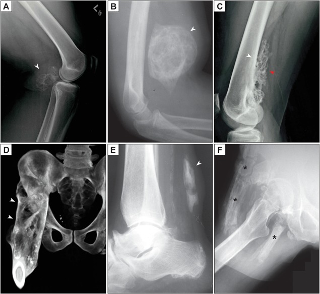
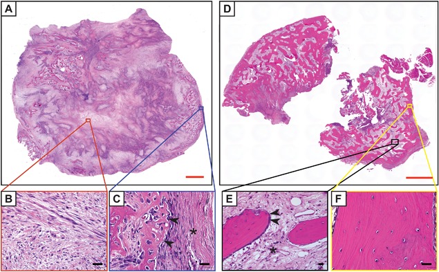
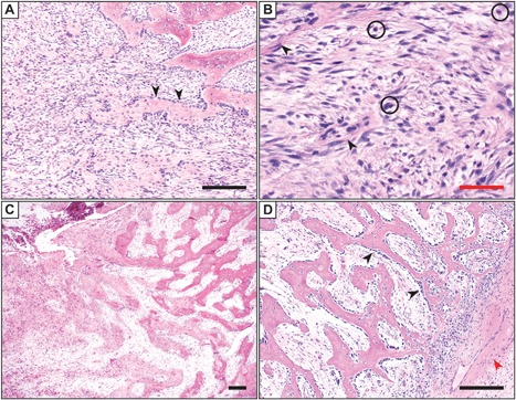
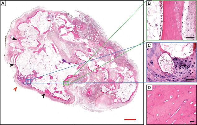
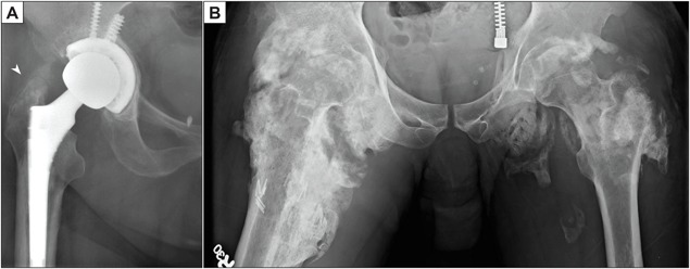
Heterotopic ossification (HO) is a diverse pathologic process, defined as the formation of extraskeletal bone in muscle and soft tissues. HO can be conceptualized as a tissue repair process gone awry and is a common complication of trauma and surgery. This comprehensive review seeks to synthesize the clinical, pathoetiologic, and basic biologic features of HO, including nongenetic and genetic forms. First, the clinical features, radiographic appearance, histopathologic diagnosis, and current methods of treatment are discussed. Next, current concepts regarding the mechanistic bases for HO are discussed, including the putative cell types responsible for HO formation, the inflammatory milieu and other prerequisite "niche" factors for HO initiation and propagation, and currently available animal models for the study of HO of this common and potentially devastating condition. © 2019 The Authors. JBMR Plus published by Wiley Periodicals, Inc. on behalf of American Society for Bone and Mineral Research.

Keywords: ECTOPIC BONE; FIBRODYSPLASIA OSSIFICANS PROGRESSIVA; HETEROTOPIC BONE; MYOSITIS OSSIFICANS.


Figures

Figure 1
Figure 1
Radiographic appearance of heterotopic ossification (HO). (A) Lateral radiograph of the knee, demonstrating a posterior, well‐circumscribed soft tissue mass with peripheral radiodensity characteristic of HO (white arrowhead). A history of blunt trauma to the area was provided. (B) Lateral radiograph of the elbow, demonstrating an anterior, well‐circumscribed, and heavily ossified soft tissue mass (white arrowhead). Peripheral radiodensity is less apparent in this example. A history of antecedent local trauma was present. (C) Later, HO may form focal or even more diffuse connections to the underlying bone. In this unusual case, a well‐demarcated osseous lesion (red arrowhead) has a broad base of attachment (white arrowhead) to the posterior aspect of the distal femur. This lesion came to clinical attention due to local discomfort but without a history of trauma. (D) In severe cases, complete fusion (ankylosis) of the joint is observed, as is the case in the CT reconstruction with broad‐based connections of bone from the proximal femur to the ilium (white arrowheads). Clinical data not available. (E) Lateral radiograph of the ankle, demonstrating ossification of the Achilles tendon (white arrowhead). A history of local trauma was provided. (F) In this case of genetic heterotopic bone (fibrodysplasia ossificans progressiva), extensive ossification (asterisks) is present within the subcutis, muscle, and deep soft tissue around the proximal femur.
Figure 2
Figure 2
Histologic appearance of heterotopic ossification (HO) by H&E staining. (A) Whole‐mount, tile scan image of early HO, demonstrating a highly cellular lesion with multifocal areas of wispy osteoid formation. This enlarging intramuscular lesion was removed from the back of a 15‐year‐old patient, with no known history of local trauma. (B) Representative central area, which demonstrates a “granulation tissue‐like” spindle cell proliferation without bone. Magnification ×40. (C) Representative peripheral area, demonstrating woven bone (arrowheads) and compressed fibrous pseudocapsule on the lesions’ exterior (asterisk). Magnification ×40. (D) Mature HO, often received in fragments owing to the density of the bone tissue. Whole‐mount, tile scan image. Here, increased quantity of thickened, lamellar bone is observed. Longstanding intramuscular mass was excised from the forearm of a 31‐year‐old patient. No history of trauma or other predisposing factor to HO was obtained in this case. (E) Representative central area, with lamellar bone trabeculae with bone‐lining cells (arrowheads) with intervening fibrous tissue resembling fibrotic marrow (asterisk). Magnification ×20. (F) Representative peripheral area, with thickened lamellar bone bearing resemblance to native cortical bone. Magnification ×40. Red scale bars = 2 mm. Black scale bars = 25 µm.
Figure 3
Figure 3
Histologic appearance of early/evolving heterotopic ossification (HO) by H&E staining. (A) The lesion is highly cellular and composed of spindle cells and scattered inflammatory cells. In the upper right, heterotopic bone formation is observed, with prominent osteoblast rimming (arrowheads). Note the intramembranous origin of bone in this case, which forms directly from stromal cell condensates. Magnification ×10. (B) Higher magnifications demonstrate the spindle cell proliferation in areas without bone formation. A cellular proliferation of spindled to ovoid cells are set in a variably edematous to fibrous stroma, with slender and elongated capillary‐type vessels (arrowheads) and a background of inflammatory cells (circles). No frank nuclear atypia or atypical mitotic figures are found. Magnification ×40. (C) A gradual continuum of woven bone (left) to more mature lamellar bone (right) is often observed in cases of nongenetic HO, which in an extraskeletal location is pathognomonic for HO. Magnification ×4. (D) Prominent bone lining osteoblasts are found (black arrowheads) in this example of HO. Also present in this image is the fibrous capsule around the periphery of the lesion (lower right), which often houses thick‐walled feeder vessels (red arrowhead). Magnification ×10. Images from parts A, B, and D are taken from the same case: a 6‐year‐old patient with an intramuscular pelvic mass and no additional clinical history. Images from C are from a 12‐year‐old patient with an intramuscular paraspinal mass. No additional clinical history available. Black scale bar = 100 µm. Red scale bar = 25 µm.
Figure 4
Figure 4
Histologic appearance of mature/late‐stage heterotopic ossification HO by H&E staining. (A) Appearance of mature HO, with thickened peripheral bone (black arrowheads), interior fatty marrow cavity (black asterisk), and foci of endochondral ossification (red arrowhead). (B) A neo‐cortex develops with elements resembling native cortical bone. Note an adipocyte‐rich interior on the left‐hand side of the image. (C) Focal metaplastic cartilage and endochondral ossification within this case are present. (D) In less common instances, sclerotic bone may develop, as seen in an osteoma. Images from A–C obtained from a 27‐year‐old patient with ossified soft tissue mass of the foot, with history of antecedent trauma; D obtained from a 66‐year‐old patient with an ossified soft tissue mass of the foot. Antecedent trauma as well as a history of previous resections was provided. All prior resections contained heterotopic bone. Red scale bars = 2 mm. Black scale bars = 50 µm.
Figure 5
Figure 5
Examples of HO post arthroplasty or spinal cord injury (neurogenic HO). (A) Flocculent radiodensity representing developing HO superior to the greater trochanter (arrowhead) in a post‐arthroplasty patient. (B) Massive and bilateral, peri‐articular HO in an immobilized patient with spinal cord injury.

References

    1. Dorfman HD, Czerniak B. Bone tumors: Maryland Heights, MO: Mosby; 1998.
    1. Ackerman LV. Extra‐osseous localized non‐neoplastic bone and cartilage formation (so‐called myositis ossificans): clinical and pathological confusion with malignant neoplasms. J Bone Joint Surg Am. 1958;40‐A(2):279–98. - PubMed
    1. de Silva MV, Reid R. Myositis ossificans and fibroosseous pseudotumor of digits: a clinicopathological review of 64 cases with emphasis on diagnostic pitfalls. Int J Surg Pathol. 2003;11 (3):187–95. - PubMed
    1. Nuovo MA, Norman A, Chumas J, Ackerman LV. Myositis ossificans with atypical clinical, radiographic, or pathologic findings: a review of 23 cases. Skeletal Radiol. 1992;21 (2):87–101. - PubMed
    1. Rosenberg AE. Pseudosarcomas of soft tissue. Arch Pathol Lab Med. 2008;132(4):579–86. - PubMed

LinkOut - more resources