Review
. 2019 Sep;7(5):631-643.
doi: 10.1111/andr.12632. Epub 2019 May 1.

Epithelial dynamics in the epididymis: role in the maturation, protection, and storage of spermatozoa

Affiliations
Review

Epithelial dynamics in the epididymis: role in the maturation, protection, and storage of spermatozoa

S Breton et al. Andrology. 2019 Sep.

Abstract

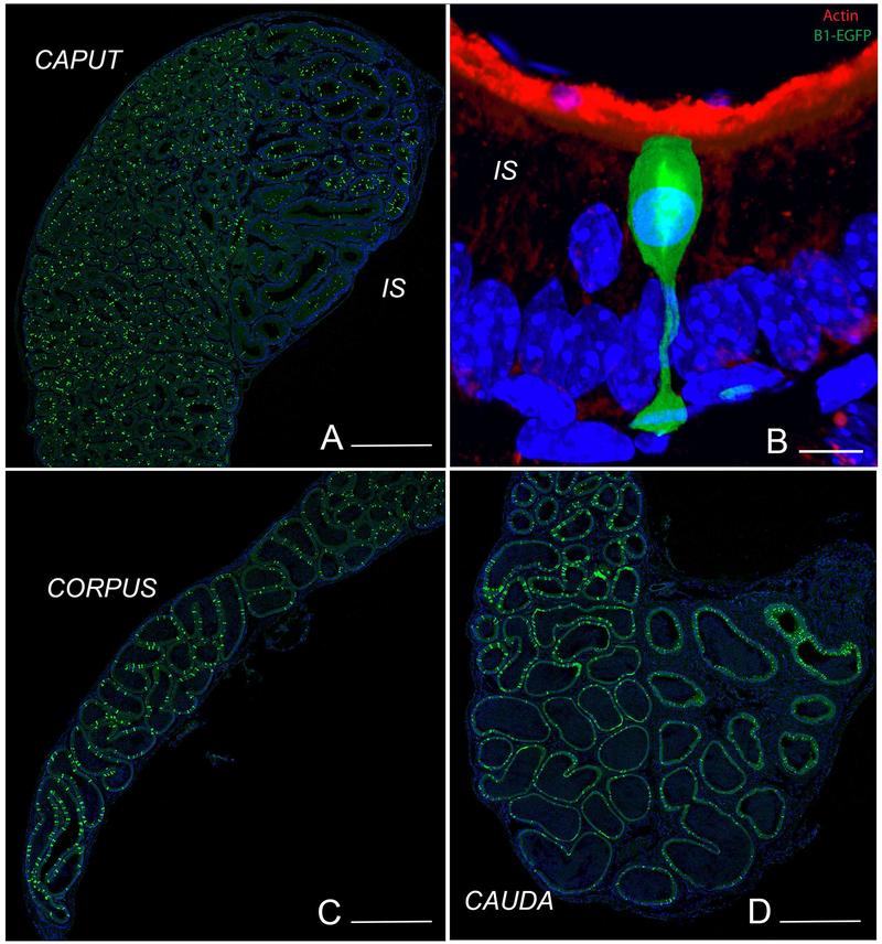
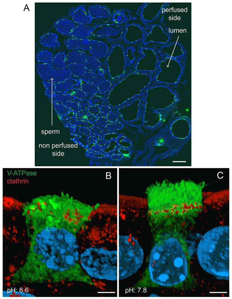
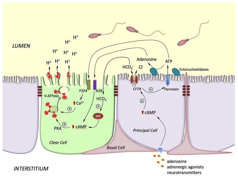
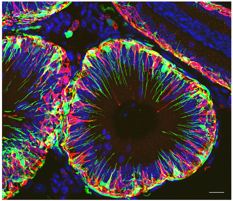
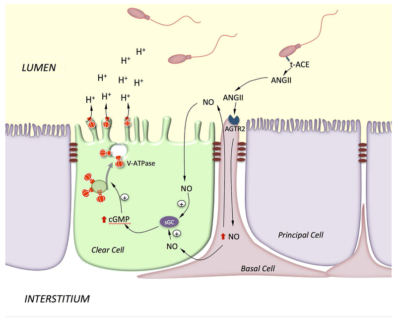
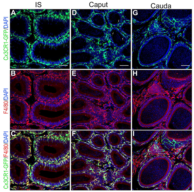
Epithelial cells line the lumen of tubular organs and are key players in their respective functions. They establish a unique luminal environment by providing a protective barrier and by performing vectorial transport of ions, nutrients, solutes, proteins, and water. Complex intercellular communication networks, specific for each organ, ensure their interaction with adjacent epithelial and non-epithelial cells, allowing them to respond to and modulate their immediate environment. In the epididymis, several epithelial cell types work in a concerted manner to establish a luminal acidic milieu that is essential for the post-testicular maturation and storage of spermatozoa. The epididymis also prevents autoimmune responses against auto-antigenic spermatozoa, while ensuring protection against ascending and blood pathogens. This is achieved by a network of immune cells that are in close contact and interact with epithelial cells. This review highlights the coordinated interactions between spermatozoa, basal cells, principal cells, narrow cells, clear cells, and immune cells that contribute to the maturation, protection, selection, and storage of spermatozoa in the lumen of the epididymis.

Keywords: epididymal immunology; luminal acidification; male fertility; post-testicular regulation; tight junctions.


Conflict of interest statement

CONFLICT OF INTEREST

SB has a financial interest in Kantum Pharma a company developing a diagnostic and therapeutic combination to prevent and treat Acute Kidney Injury. SB’s interests were reviewed and are managed by Massachusetts General Hospital and Partners HealthCare in accordance with their conflict of interest policies.

Figures

Figure 1)
Figure 1). Visualization of NCs and CCs in the epididymis of transgenic mice expressing EGFP under the control of the promoter of the V-ATPase B1 subunit (B1-EGFP).
EGFP+ NCs (green) are located in the IS, and EGFP+ CCs (green) are located in the caput (A), corpus (C) and cauda (D) regions. B) In the IS, NCs have a “champagne glass” appearance and their nuclei are located in the apical region of the epithelium, compared to adjacent PCs. A dense network of filamentous actin is seen in the apical stereocilia of PCs (labeled in red using phalloidin). Nuclei are labeled in blue using DAPI. Bars: A, C, D = 500 μm, B = 5 μm.
Figure 2)
Figure 2). Effect of luminal pH on V-ATPase sub-cellular localization in CCs in the cauda epididymis.
Mouse cauda epididymidis was perfused in vivo, and cryostat sections of fixed tissues were stained for the V-ATPase B1 subunit (green). Nuclei are labeled in blue with DAPI. A) Luminal spermatozoa are absent from the perfused tubules. Luminal V-ATPase positive CCs are detected in the perfused and non-perfused regions. B) 3D reconstruction by Airyscan microscopy of a CC perfused with a solution adjusted to the control pH of 6.6. Labeling for the endocytic marker clathrin (red) was performed to identify the apical border of the cell. Short V-ATPase-labeled microplicae and intracellular V-ATPase labeling are observed in this “resting” cell (green). Adjacent PCs are seen with intracellular clathrin labeling. C) 3D reconstruction by Airyscan microscopy of a CC perfused with a solution adjusted to the alkaline pH of 7.8. Double-labeling for V-ATPase (green) and clathrin (red) was performed. Numerous long V-ATPase-labeled microplicae are observed in this “activated” cell, and less intracellular V-ATPase labeling is detected (green). Adjacent PCs are seen with intracellular clathrin labeling. Bars: A = 400 μm; B, C = 2.5 μm. (Modified from (Battistone et al., 2019).
Figure 3)
Figure 3). Model showing the coordinated interaction between PCs and CCs for the activation of V-ATPase-dependent proton secretion in CCs.
Stimulation of PCs by basolateral factors such as adenosine, adrenergic agonists or neurotransmitters elevate intracellular cAMP, which in turns activates bicarbonate secretion via apical CFTR. Luminal bicarbonate then reaches CCs, where it activates the soluble adenylate cyclase (sAC) to produce cAMP. CFTR also stimulates ATP secretion via pannexin activation. ATP either activates P2X4 located on the apical membrane of CCs, or is hydrolyzed by extracellular ectonucleotidases to produce adenosine. Adenosine activates the GPCR receptor ADORA2B (A2B) located on the apical membrane of CCs, leading to elevation in intracellular cAMP. Activation of the cAMP/PKA pathway by bicarbonate or adenosine induces V-ATPase apical membrane accumulation. On the other hand, activation of P2X4 by luminal ATP in CCs induces an elevation of intracellular calcium, which also facilitates V-ATPase membrane accumulation. The redistribution of V-ATPase from intracellular vesicles to the apical membrane results in the formation and elongation of V-ATPase-enriched microplicae in CCs and stimulates proton secretion.
Figure 4)
Figure 4). Confocal microscopy visualization of BCs and MPs in the IS of CD11c-EYFP transgenic mice.
Immunofluorescence labeling for the BC specific marker keratin 5 (KRT5; red) shows abundant BCs with luminal reaching axiopodia in the IS. CD11c-EYFP positive MPs (green) also send luminal reaching projections, but they are clearly distinct from BCs. Nuclei are labeled in blue with DAPI. Bar = 15 μm.
Figure 5)
Figure 5). Model showing BC-CC crosstalk via the ANGII signaling pathway.
Axiopodia in BCs cross the TJs and are in contact with the lumen. Luminal ANGII stimulates AGTR2 in BCs and induces the production of NO, which then diffuses out to reach CCs, where it produces cGMP via activation of soluble guanylate cyclase (sGC). cGMP triggers V-ATPase apical accumulation and increases proton secretion by CCs. ANGII is produced by testicular ACE (t-ACE), which is attached to the sperm membrane via a GPI linker.
Figure 6)
Figure 6). Confocal microscopy showing a dense network of mononuclear phagocytes in all epididymal segments.
Epididymis of CX3CR1-EGFP transgenic mice was labeled for the macrophage marker F4/80 (red). A,B,C) In the IS, most CX3CR1 positive MPs (green) are also labeled for F4/80 identifying them as macrophages. The majority of macrophages are in close proximity with the epithelium and show numerous intraepithelial projections extending toward the lumen. D,E,F) In the caput, most CX3CR1 positive MPs (green) are also labeled for F4/80 identifying them as macrophages. These macrophages are in close proximity with the epithelium but only a few rare cells now send intraluminal projections towards the lumen (arrows in D). A significant number of cells positive for both CX3CR1 and F4/80 are located in the interstitium. G, H, I) In the cauda, cells positive for CX3CR1 and F4/80 are located next to the epithelium but they do not extend intraepithelial projections. In the interstitium, a mixed population of cells double positive for CX3CR1 and F4/80, and cells positive for F4/80 but negative for CX3CR1 are detected. Nuclei are labeled in blue with DAPI.

Similar articles

Cited by

References

    1. Andrews C, McLean MH, Durum SK. Cytokine Tuning of Intestinal Epithelial Function. Front Immunol 2018;9:1270. - PMC - PubMed
    1. Au CL, Wong PY. Luminal acidification by the perfused rat cauda epididymidis. J Physiol 1980;309:419–27. - PMC - PubMed
    1. Bagnis C, Marsolais M, Biemesderfer D, Laprade R, Breton S. Na+/H+-exchange activity and immunolocalization of NHE3 in rat epididymis. Am J Physiol Renal Physiol 2001;280:F426–36. - PubMed
    1. Balda MS, Matter K. The tight junction protein ZO-1 and an interacting transcription factor regulate ErbB-2 expression. EMBO J 2000;19:2024–33. - PMC - PubMed
    1. Banks FC, Calvert RC, Burnstock G. Changing P2X receptor localization on maturing sperm in the epididymides of mice, hamsters, rats, and humans: a preliminary study. Fertil Steril 2010;93:1415–20. - PubMed

Publication types

LinkOut - more resources