. 2019 Aug;35(8):710-717.
doi: 10.1089/AID.2019.0039. Epub 2019 May 29.

HIV Transcription Is Independent of Mediator Kinases

Affiliations

HIV Transcription Is Independent of Mediator Kinases

Daniele C Cary et al. AIDS Res Hum Retroviruses. 2019 Aug.

Abstract

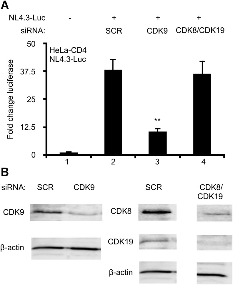
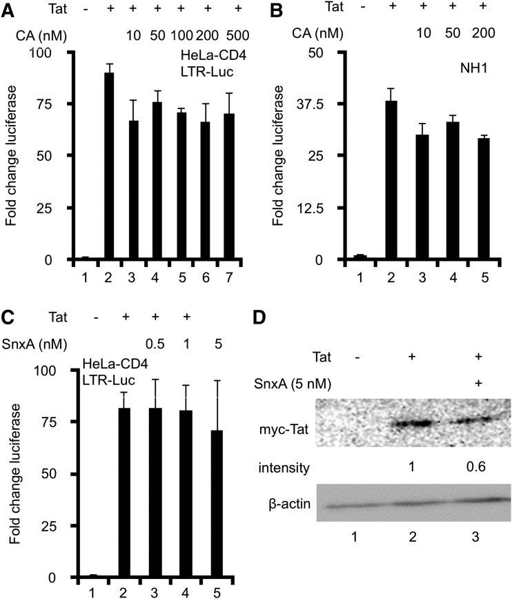
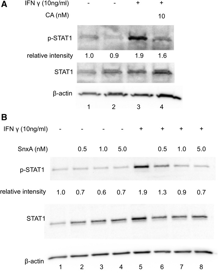
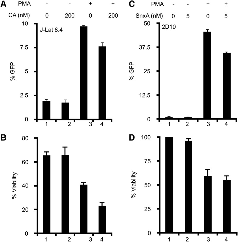
While the roles in HIV transcription of many cyclin-dependent kinases (CDKs) have been well defined, little is known about the impact of mediator kinases (MDKs), CDK8 and CDK19, in this process. Mediator complexes containing CDK8 or CDK19 repress or activate the expression of selected genes. The aim of this study was to investigate the role of MDKs in HIV transcription. siRNA knockdown of both MDKs had no effect on HIV transcription. This result was confirmed using two MDK inhibitors, Cortistatin A (CA) and Senexin A (SnxA). Furthermore, neither CA nor SnxA inhibited viral reactivation in Jurkat cell models of HIV latency. Taken together, these results indicate that MDKs are not required for HIV transcription.

Keywords: CDK19; CDK8; Cortistatin A; HIV transcription; Senexin A; mediator kinases.


Conflict of interest statement

No competing financial interests exist.

Figures

<b>FIG. 1.</b>
FIG. 1.
Knockdowns of MDKs do not inhibit HIV transcription. HeLa-CD4 cells were transfected with siRNAs against CDK9, CDK8, and CDK19, or scrambled non-specific control (SCR). Twenty-four hours post knockdown, cells were transfected with NL4.3 Luc. At 24 h post NL4.3-Luc transfection (48 h post knockdown), lysates were harvested and examined for luciferase activity or Western blotting. (A) Luciferase activity is presented as a fold change in luciferase activity over untreated control cells (set to 1). (B) Whole-cell lysates were run on 10% SDS-PAGE and blotted with anti-CDK9, anti-CDK8, anti-CDK19 antibodies, with β-actin serving as loading control. Samples from cells treated with scrambled and CDK8/19 siRNAs were run on the same gel. Triplicate stimulations were performed. Error bars represent standard errors of the mean (**p < .01). CDK, cyclin-dependent kinase; MDK, mediator kinase.
<b>FIG. 2.</b>
FIG. 2.
Chemical inhibition of MDKs does not inhibit HIV transcription. (A) HeLa-CD4 cells were co-transfected with LTR-Luc and Tat for 48 h. CA was added at the indicated concentrations. Luciferase activity was measured 48 h post transfection, and is presented as a fold change in luciferase activity over untreated control cells (set to 1). (B) NH1 cells, stably expressing LTR-Luc, were transfected with Tat ± CA for 48 h. Luciferase activity was measured 48 h post transfection. (C) HeLa-CD4 cells were co-transfected with LTR-Luc and Tat for 48 h. SnxA was added at indicated concentrations. Luciferase activity was measured 48 h post transfection. (D) Whole-cell lysates of SnxA-treated HeLa-CD4 cells were separated on 15% SDS PAGE. The membrane was probed with anti-myc and anti-β-actin antibodies. Densitometry was calculated and normalized to β-actin. Results are representative of two independent experiments. Triplicate stimulations were performed for all experiments. Error bars represent standard errors of the mean. CA, Cortistatin A; LTR-Luc, long-terminal repeat luciferase; SnxA, Senexin A.
<b>FIG. 3.</b>
FIG. 3.
Chemical inhibition of MDKs decreases STAT1 phosphorylation. HeLa-CD4 cells were pretreated with (A) 10 nM CA for 1 h or (B) indicated concentrations of SnxA, then stimulated with IFNγ (10 ng/mL) for 1 h. Whole-cell lysates were run on 10% SDS-PAGE and blotted with anti-phospho-STAT1 and anti-STAT1 antibodies with β-actin serving as loading control. Representative blots of triplicate stimulations are presented. IFNγ, interferon-gamma.
<b>FIG. 4.</b>
FIG. 4.
Chemical inhibition of MDKs does not inhibit HIV reactivation. (A) J-Lat 8.4 cells were stimulated with PMA (12 nM) ± CA (200 nM) for 24 h. GFP expression of viable cells was measured by flow cytometry. (B) Percent viability was estimated using the percentage of live lymphocytes in 10,000 total cells analyzed. Untreated control cell viability was set at 100% viability. (C) 2D10 cells were stimulated with PMA (12 nM) ± SnxA (5 nM) for 24 h. GFP expression of viable cells was measured by flow cytometry. (D) Percent viability was estimated using the percentage of live lymphocytes in 10,000 total cells analyzed. Untreated control cell viability was set at 100% viability. Triplicate stimulations were performed. Error bars represent standard errors of the mean. GFP, green fluorescent protein; PMA, phorbol myristate acetate.

Similar articles

Cited by

References

    1. Cary DC, Fujinaga K, Peterlin BM: Molecular mechanisms of HIV latency. J Clin Invest 2016;126:448–454 - PMC - PubMed
    1. Cary DC, Fujinaga K, Peterlin BM: Euphorbia Kansui Reactivates Latent HIV. PLoS One 2016;11:e0168027. - PMC - PubMed
    1. Bartholomeeusen K, Fujinaga K, Xiang Y, Peterlin BM: Histone deacetylase inhibitors (HDACis) that release the positive transcription elongation factor b (P-TEFb) from its inhibitory complex also activate HIV transcription. J Biol Chem 2013;288:14400–14407 - PMC - PubMed
    1. Cujec TP, Okamoto H, Fujinaga K, et al. : The HIV transactivator TAT binds to the CDK-activating kinase and activates the phosphorylation of the carboxy-terminal domain of RNA polymerase II. Genes Dev 1997;11:2645–2657 - PMC - PubMed
    1. Ivanov A, Lin X, Ammosova T, et al. : HIV-1 Tat phosphorylation on Ser-16 residue modulates HIV-1 transcription. Retrovirology 2018;15:39. - PMC - PubMed

Publication types

MeSH terms

LinkOut - more resources