

Myocardial fibrosis by late gadolinium enhancement cardiovascular magnetic resonance in myotonic muscular dystrophy type 1: highly prevalent but not associated with surface conduction abnormality
- PMID: 31046780
- PMCID: PMC6498496
- DOI: 10.1186/s12968-019-0535-6
Myocardial fibrosis by late gadolinium enhancement cardiovascular magnetic resonance in myotonic muscular dystrophy type 1: highly prevalent but not associated with surface conduction abnormality
Abstract
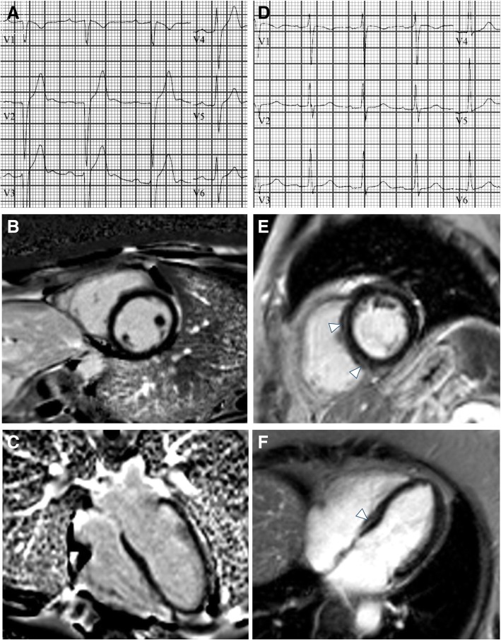
Background: Conduction disease and arrhythmias represent a major cause of mortality in myotonic muscular dystrophy type 1 (MMD1). Permanent pacemaker (PPM) implantation is the cornerstone of therapy to reduce cardiovascular mortality in MMD1. Cardiovascular magnetic resonance (CMR) studies demonstrate a high prevalence of myocardial fibrosis in MMD1, however the association between CMR myocardial fibrosis with late gadolinium enhancement (CMR-LGE) and surface conduction abnormality is not well established in MMD1. We investigated whether myocardial fibrosis by CMR-LGE is associated with surface conduction abnormalities meeting criteria for PPM implantation according to current guidelines in a cohort of patients with genetically confirmed MMD1.
Methods: Patients with genetically confirmed MMD1 were retrospectively evaluated. 12-lead electrocardiography (ECG) performed within 6 months of CMR was necessary for inclusion. The severity and extent of MMD1 was quantified using a validated Muscular Impairment Rating Scale (MIRS). Based on current guidelines for device-based therapy of cardiac rhythm abnormalities, we defined surface conduction abnormality as the presence of ECG alterations meeting criteria for PPM implant (class I or II indications): PR interval > 200 ms (type I atrioventricular (AV) block) and/or mono or bifascicular block (QRS > 120 ms), or evidence of advanced AV block. Balanced steady-state free precession sequences (bSSFP) were used for assessment of left ventricular (LV) volumes and ejection fraction. MOdified Look-Locker Inversion Recovery (MOLLI) acquisition schemes were used to acquire T1 maps. Patients' charts were reviewed up to 12 months post-CMR for occurrence of PPM implantation.
Results: Fifty-two patients (38% male, 41 ± 14 years) were included. Overall, 31 (60%) patients had a surface conduction abnormality and 22 (42%) demonstrated midwall myocardial fibrosis by CMR-LGE. After a median of 57 days from CMR exam, 15 patients (29%) underwent PPM implantation. Subjects with vs. without surface conduction abnormality had significantly longer disease length (15.5 vs. 7.8 years, p = 0.015) and higher disease severity on the MIRS scale (p = 0.041). High prevalence of myocardial fibrosis by CMR-LGE was detected in subjects with and without surface conduction abnormality with no significant difference between the two cohorts (42% vs. 43%, p = 0.999). By multivariate logistic regression analysis, disease length was the only independent variable associated with surface conduction abnormality (OR 1.071, 95%CI 1.003-1.144, p = 0.040); while CMR-LGE was not associated with conduction abnormality (ρ = - 0.009, p = 0.949).
Conclusions: Myocardial fibrosis by CMR-LGE is highly prevalent in MMD1 but not related to surface conduction abnormality meeting current guideline criteria for PPM implantation .
Keywords: Cardiovascular magnetic resonance; Electrocardiogram; Late gadolinium enhancement; Myocardial fibrosis; Myotonic muscular dystrophy; Pacemaker.
Conflict of interest statement
Authors’ information
No third parties were involved in the authorship of review of the manuscript.
Consent for publication
Not applicable. All data presented in this article is non-identifiable.
Competing interests
The authors have declared that they have no competing interests.
Publisher’s Note
Springer Nature remains neutral with regard to jurisdictional claims in published maps and institutional affiliations.
Figures
References
-
- Epstein AE, DiMarco JP, Ellenbogen KA, et al. 2012 ACCF/AHA/HRS focused update incorporated into the ACCF/AHA/HRS 2008 guidelines for device-based therapy of cardiac rhythm abnormalities: a report of the American College of Cardiology Foundation/American Heart Association Task Force on Practice Guidelines and the Heart Rhythm Society. J Am Coll Cardiol. 2013;61:e6–75. doi: 10.1016/j.jacc.2012.12.014. - DOI - PubMed
Publication types
MeSH terms
Substances
Grants and funding
LinkOut - more resources
Full Text Sources
Medical
Research Materials
