

Multiple spindle cell hemangiomas in both lungs: a rare case report and review of the literature
- PMID: 31046794
- PMCID: PMC6498687
- DOI: 10.1186/s13019-019-0906-y
Multiple spindle cell hemangiomas in both lungs: a rare case report and review of the literature
Abstract
Background: Spindle cell hemangioma (SCH) was an extremely rare benign tumor which typically arised in the subcutis of the distal extremities of young people. In this study, we reported a case of multiple spindle cell hemangioma in both lungs.
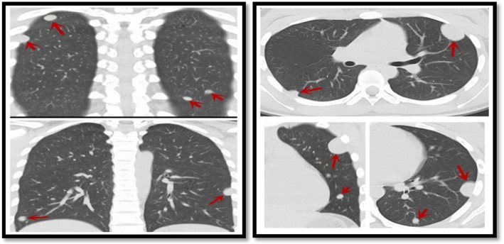
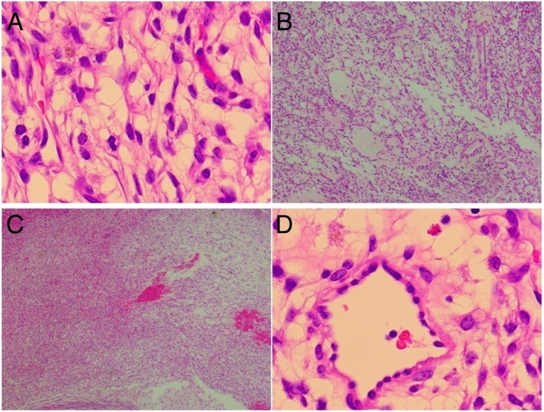
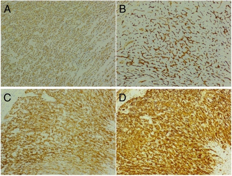
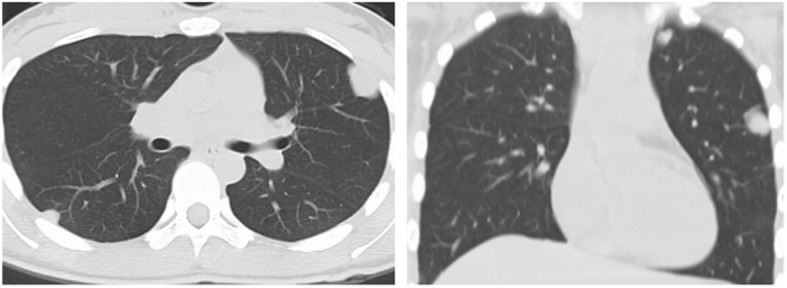
Case presentation: A 19-year-old HIV-negative female was found to have multiple lung nodules by the chest X-ray during the physical examination. Her chest CT scan revealed multiple round-like pulmonary nodules in both lungs. Based on the morphological features and immunohistochemical examination for vascular markers CD31, CD34 and D2-40, the mass was diagnosed as SCH after surgery.
Conclusion: SCH was an extremely rare tumor especially in both lungs. It should be considered in differential diagnosis of multiple lung nodules. Pathological features, the expression of CD31, CD34 and D2-40 could help to diagnosis of SCH.
Keywords: Hemangioma of lung; Multiple nodules of lung; Spindle cell hemangiomas.
Conflict of interest statement
Ethics approval and consent to participate
Not applicable.
Consent for publication
Consent for publication of this case report in its entirety was obtained from the patient.
Competing interests
The authors declare that they have no competing interests.
Publisher’s Note
Springer Nature remains neutral with regard to jurisdictional claims in published maps and institutional affiliations.
Figures

References
Publication types
MeSH terms
LinkOut - more resources
Full Text Sources
Medical
Miscellaneous
