Review
. 2019 Aug:200:126-134.
doi: 10.1016/j.pharmthera.2019.04.010. Epub 2019 Apr 29.

Signaling in the microenvironment of pancreatic cancer: Transmitting along the nerve

Affiliations
Review

Signaling in the microenvironment of pancreatic cancer: Transmitting along the nerve

Noelle Jurcak et al. Pharmacol Ther. 2019 Aug.

Abstract

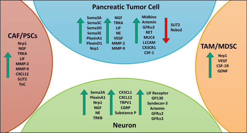
Pancreatic ductal adenocarcinoma (PDA) is a dismal malignant disease with the lowest stage-combined overall survival rate compared to any other cancer type. PDA has a unique tumor microenvironment (TME) comprised of a dense desmoplastic reaction comprising over two-thirds of the total tumor volume. The TME is comprised of cellular and acellular components that all orchestrate different signaling mechanisms together to promote tumorigenesis and disease progression. Particularly, the neural portion of the TME has recently been appreciated in PDA progression. Neural remodeling and perineural invasion (PNI), the neoplastic invasion of tumor cells into nerves, are common adverse histological characteristics of PDA associated with a worsened prognosis and increased cancer aggressiveness. The TME undergoes dramatic neural hypertrophy and increased neural density that is associated with many signaling pathways to promote cell invasion. PNI is also considered one of the main routes for cancer recurrence and metastasis after surgical resection, which remains the only current cure for PDA. Recent studies have shown multiple cell types in the TME signal through autocrine and paracrine mechanisms to enhance perineural invasion, pancreatic neural remodeling and disease progression in PDA. This review summarizes the current findings of the signaling mechanisms and cellular and molecular players involved in neural signaling in the TME of PDA.

Keywords: Neural remodeling; Pancreatic ductal adenocarcinoma; Perineural invasion; Tumor microenvironment.


Conflict of interest statement

Conflict of Interest:

The authors have no relevant conflict of interests to disclose.

Figures

Figure 1
Figure 1. Signaling interactions impacting neural architecture and PNI in PDA.
There are several complex paracrine signaling interactions that lead to the dramatic changes in neural architecture during PDA development and progression. Tumor cells reciprocally signal with neurons, CAF/PSCs, and TAM/MDSCs through axon guidance, cytokine and NGF signaling to promote changes in ECM remodeling. In addition, tumor cells and neurons utilize catecholamine signaling in the TME. Comprehensively, these signaling pathways increase the TME capability for neuron remodeling, neuron outgrowth, perineural invasion and tumor growth and metastasis in PDA. Abbreviations: ECM-Extracellular matrix, CAF-Cancer-associated fibroblast, PSC-Pancreatic Stellate Cell, NGF-Neural growth factor, TAM-Tumor-associated macrophage, and MDSC-Myeloid derived suppressor cell.
Figure 2
Figure 2. Major molecular players involved in neural signaling in the TME.
During PDA tumorigenesis and disease progression the expression of several molecular factors is dysregulated. This figure highlights some of the major molecular players that have been identified in increasing PDA cell invasion, perineural invasion and neuron remodeling in the cell types of the PDA TME. The green arrows indicate molecular player that have increased expression in PDA and the red arrow indicates molecular players with decreased expression in PDA.

References

    1. Ahrens D Von Bhagat TD, Nagrath D, Maitra A, & Verma A (2017). The role of stromal cancer-associated fibroblasts in pancreatic cancer. Journal of Hematology & Oncology, 10(76), 1–8. doi:10.1186/s13045-017-0448-5. - DOI - PMC - PubMed
    1. Alrawashdeh W, Jones R, Dumartin L, Radon TP, Cutillas PR, Feakins RM, … Crnogorac‐Jurcevic T (2019). Perineural invasion in pancreatic cancer: proteomic analysis and in vitromodelling. Molecular Oncology, 10.1002/1878-0261.12463. - DOI - PMC - PubMed
    1. Amit M, Na’Ara S, & Gil Z (2016). Mechanisms of cancer dissemination along nerves. Nature Reviews Cancer, 16(6), 399–408. doi:10.1038/nrc.2016.38. - DOI - PubMed
    1. Amit M, Na S, Binenbaum Y, Kulish N, Fridman E, Shabtai-Orbach A, … Gil Z (2017). Upregulation of RET induces perineurial invasion of pancreatic adenocarcinoma. Oncogene, 36(23), 3232–3239. doi:10.1038/onc.2016.483. - DOI - PubMed
    1. Bakst RL, Xiong H, Chen C, Deborde S, Lyubchik A, Zhou Y, … Wong RJ (2018). Inflammatory monocytes promote perineural invasion via CCL2-mediated recruitment and cathepsin B expression. Cancer Research, 77(22), 6400–6414. doi:10.1158/0008-5472.CAN17-1612. - DOI - PMC - PubMed

Publication types