. 2019 Jun 3;151(6):834-849.
doi: 10.1085/jgp.201812143. Epub 2019 May 2.

Agonists that stimulate secretion promote the recruitment of CFTR into membrane lipid microdomains

Affiliations

Agonists that stimulate secretion promote the recruitment of CFTR into membrane lipid microdomains

Asmahan Abu-Arish et al. J Gen Physiol. .

Abstract

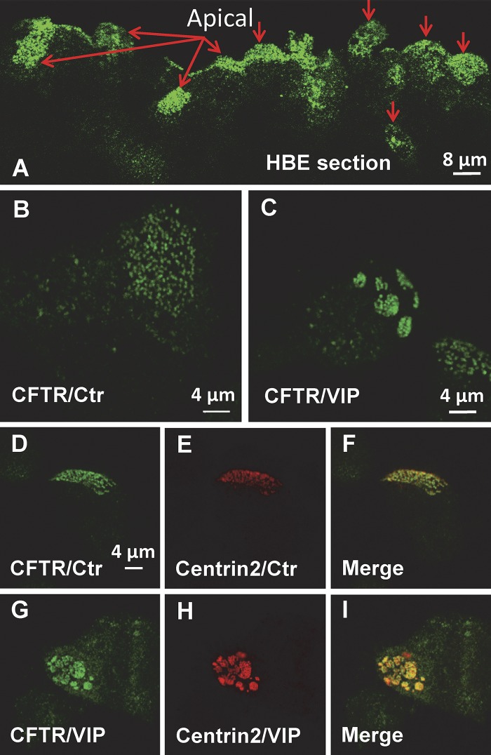
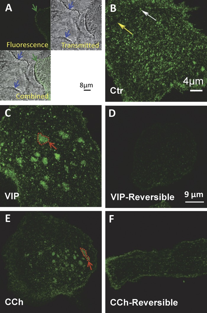
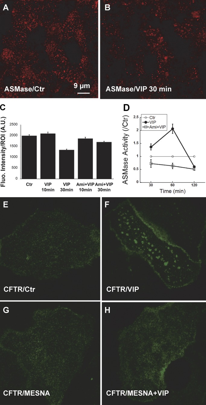
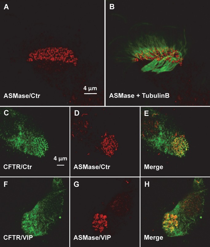
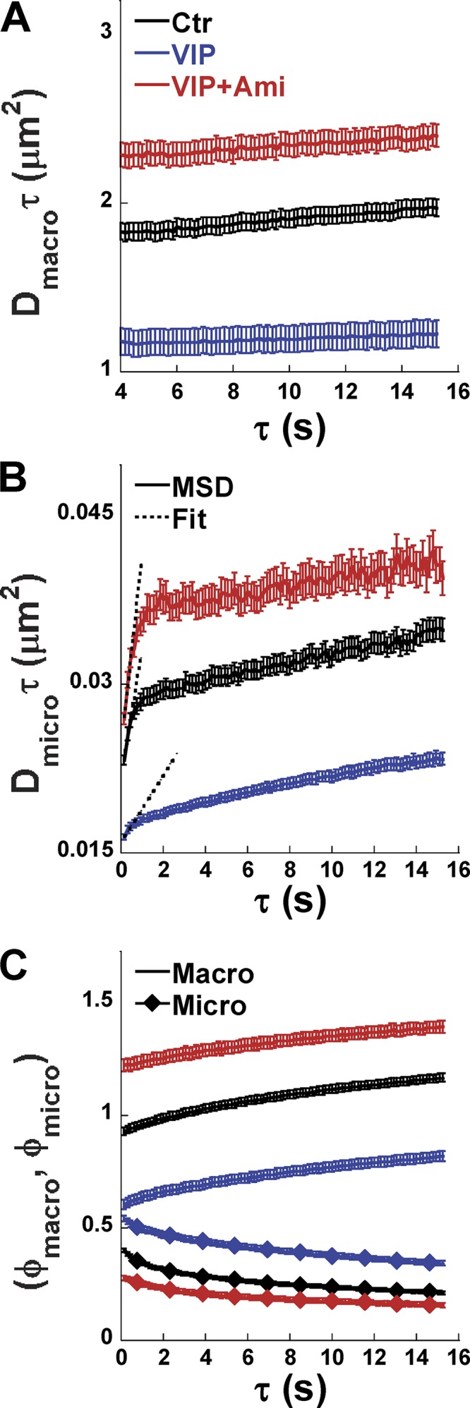
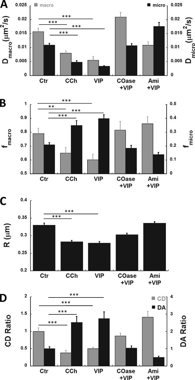
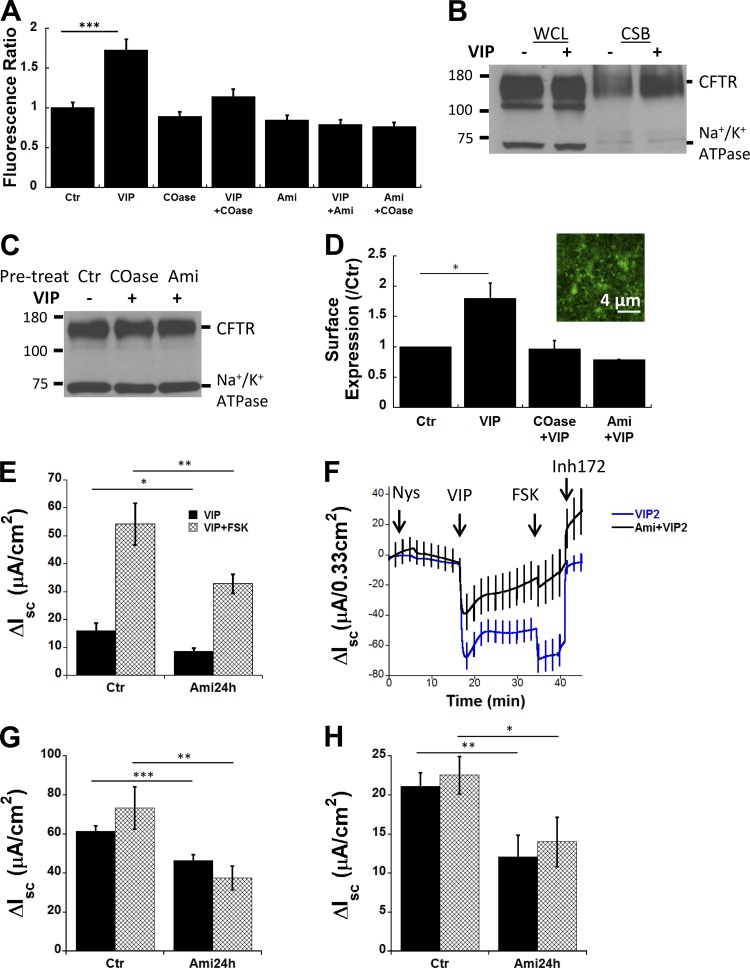
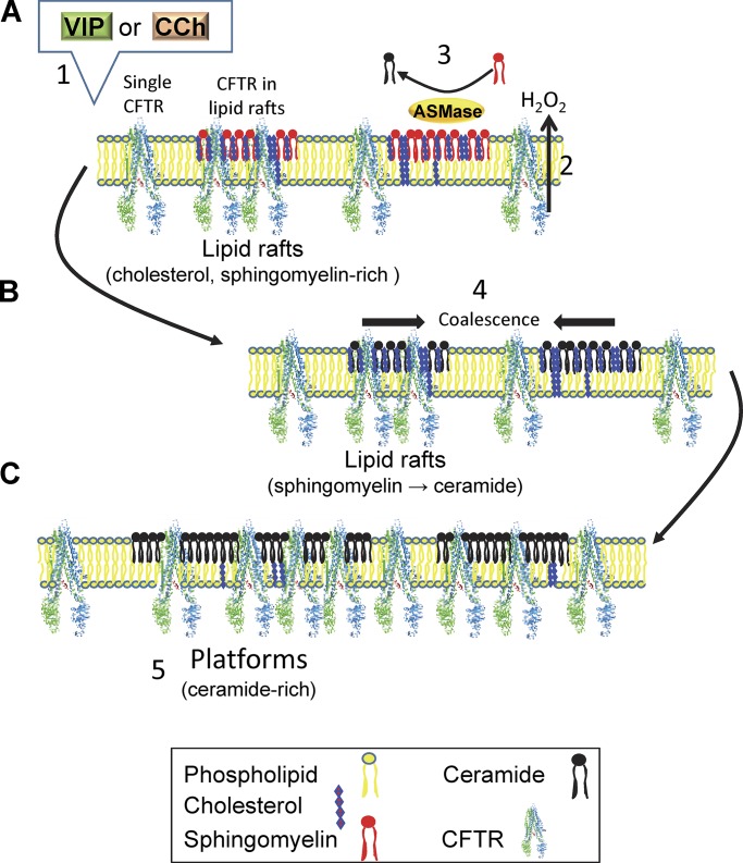
The cystic fibrosis transmembrane conductance regulator (CFTR) is a tightly regulated anion channel that mediates secretion by epithelia and is mutated in the disease cystic fibrosis. CFTR forms macromolecular complexes with many proteins; however, little is known regarding its associations with membrane lipids or the regulation of its distribution and mobility at the cell surface. We report here that secretagogues (agonists that stimulate secretion) such as the peptide hormone vasoactive intestinal peptide (VIP) and muscarinic agonist carbachol increase CFTR aggregation into cholesterol-dependent clusters, reduce CFTR lateral mobility within and between membrane microdomains, and trigger the fusion of clusters into large (3.0 µm2) ceramide-rich platforms. CFTR clusters are closely associated with motile cilia and with the enzyme acid sphingomyelinase (ASMase) that is constitutively bound on the cell surface. Platform induction is prevented by pretreating cells with cholesterol oxidase to disrupt lipid rafts or by exposure to the ASMase functional inhibitor amitriptyline or the membrane-impermeant reducing agent 2-mercaptoethanesulfonate. Platforms are reversible, and their induction does not lead to an increase in apoptosis; however, blocking platform formation does prevent the increase in CFTR surface expression that normally occurs during VIP stimulation. These results demonstrate that CFTR is colocalized with motile cilia and reveal surprisingly robust regulation of CFTR distribution and lateral mobility, most likely through autocrine redox activation of extracellular ASMase. Formation of ceramide-rich platforms containing CFTR enhances transepithelial secretion and likely has other functions related to inflammation and mucosal immunity.


Figures

Figure 1.
Figure 1.
Immunolocalization of endogenous CFTR and Centrin2. Primary HBE cells were immunostained in superior bronchial tissue (A) and primary HBE cells (B–I) that had been cultured for >3 wk at the air–liquid interface. (A) Distribution of CFTR in non-CF bronchial tissue shows puncta and platforms. Red arrows point to the apical surface. (B and C) Distribution of endogenous CFTR under control (Ctr) conditions and after 1 h bilateral exposure to 200 nM VIP at 37°C, respectively. Note both diffuse and punctate immunostaining under Ctr conditions and aggregation into large (2–4 µm dia.) platforms on cells after VIP exposure. (D–F) Distribution of endogenous CFTR (CFTR/Ctr) and the ciliogenesis marker Centrin2 (Centrin2/Ctr) under Ctr conditions. Note the colocalization of CFTR puncta with cilia basal bodies. (G–I) Redistribution of endogenous CFTR and Centrin2 during VIP stimulation. Note the aggregation of CFTR and Centrin2, which remain colocalized. Images are representative of n = 20–40 cells in at least two experiments under each condition.
Figure 2.
Figure 2.
Reversible aggregation of EGFP-CFTR induced by VIP and CCh in pHBE cells. pHBE cells were transduced with EGFP-CFTR adenovirus and cultured on collagen-coated glass for 4 d. (A) Autofluorescence is negligible compared with EGFP-CFTR fluorescence. Upper left: Confocal image of pHBE cell transiently expressing EGFP-CFTR (green arrow). Upper right: Transmitted light image of the same field reveals the presence of nonfluorescent cells (blue arrows). Lower left: Combined image showing that autofluorescence of untransduced cells (blue arrow) is negligible compared with the EGFP-CFTR expressing cell (green arrow). (B) Distribution of EGFP-CFTR under Ctr conditions showing diffuse background staining (yellow arrow) and bright puncta (white arrow). (C) Aggregation of EGFP-CFTR into large (2–4 µm dia.) platforms after 20–30 min exposure to 200 nM VIP at 37°C (red arrow shows one example). The scale bar in B also applies to C. (D) Platforms have dispersed 24 h after VIP washout. (E) Aggregation of EGFP-CFTR into large platforms induced by 20–30 min exposure to 2 µM CCh at 37°C. (F) No platforms remain 24 h after CCh washout. The scale bar in D also applies to E and F.
Figure 3.
Figure 3.
Constitutive binding of ASMase on the cell surface and its redox activation by VIP. (A and B) Immunofluorescence detection of ASMase on the apical membrane of unpermeabilized, well differentiated pHBE cells under Ctr conditions (A)and 30 min after treatment with 200 nM VIP (B). (C) Mean fluorescence intensity of ROIs (1,024 × 1,024 pixels; n = 50–130 ROIs per condition, cells from four donors studied in separate experiments). Cells were imaged under Ctr conditions, after 10 or 30 min stimulation with VIP, or following 1 h pretreatment with Ami (13 µM) followed by 10 or 30 min VIP. Stimulation for 30 min reduced ASMase surface immunostaining by 33%. (D) Normalized ASMase activity at the plasma membrane of subconfluent pHBEs expressing EGFP-CFTR at 30, 60, and 120 min after treatment with VIP. Pretreating cells with 13 µM Ami for 1 h reduced ASMase activity below the Ctr level, suggesting there is residual ASMase activity under basal conditions. Mean ± SEM. (E–H) Oxidation of extracellular ASMase stimulates formation of ceramide-rich platforms. (E) Confocal images of pHBE cells show the distribution of EGFP-CFTR under Ctr conditions. (F) EGFP-CFTR aggregation into large platforms after 20 min exposure to 200 nM VIP at 37°C. (G) The distribution of CFTR in unstimulated cells is unaffected by the extracellular reducing agent MESNA (100 mM). (H) MESNA blocks the formation of platforms in response to VIP; compare with F. Fluo., fluorescence; A.U., arbitrary units.
Figure 4.
Figure 4.
Colocalization of endogenous ASMase and CFTR. (A) Punctate distribution of ASMase on the apical surface of well-differentiated, ciliated pHBEs under Ctr conditions. (B) ASMase immunofluorescence is detected at the base of each motile cilium (β-tubulin immunostaining). (C–E) Co-localization of endogenous CFTR and ASMase under Ctr conditions. (F–H) Redistribution of CFTR and ASMase into platforms during VIP stimulation.
Figure 5.
Figure 5.
VIP alters CFTR dynamics, and its effects are blocked by Ami. (A) Mean squared displacement Dmacroτ increases linearly as a function of τ under all conditions, and its slope (Dmacro) yields the effective macroscopic diffusion coefficient for the unconfined CFTR population. (B) Mean squared displacement Dmicroτ increases linearly for the first few temporal lags under all conditions, and then its slope decreases at long τ. The first three Dmicroτ values were fitted with straight lines as shown to obtain the initial slope and Dmicro, which quantifies CFTR mobility inside confinements (microdomains). VIP treatment resulted in a smaller Dmicro compared with Ctr conditions, and this decrease was abolished by pretreating cells with Ami to inhibit ceramide production. (C) Amplitudes of the macro (ϕmacro, line only) and micro (ϕmicro, diamond symbols) components of the k-space correlation function and their dependence on τ. Mean ± SEM. For the number of cells analyzed, see Table 1.
Figure 6.
Figure 6.
CFTR aggregation and dynamics are regulated by VIP and CCh through lipid-dependent mechanisms. pHBE cells transduced with EGFP-CFTR adenovirus were imaged 4 d later under Ctr conditions and during exposure to CCh or VIP alone, VIP after 1 h pretreatment with COase to disrupt rafts (COase+VIP), or VIP after 40 min pretreatment with Ami to inhibit ceramide synthesis by ASMase (Ami+VIP). (A) Both Dmacro and Dmicro of CFTR were decreased significantly after VIP or CCh treatment. This decrease was prevented by pretreating cells with COase to disrupt lipid rafts or Ami to inhibit ceramide synthesis. (B) The unconfined fraction of the CFTR population fmacro (gray bars) decreased during VIP or CCh exposure but not if cells were pretreated with COase or Ami. The confined CFTR population fraction fmicro (black bars) increased approximately twofold during VIP stimulation, and this was prevented by pretreating cells with COase or Ami, confirming the lipid dependence of CFTR redistribution. (C) VIP reduced the effective diffusion radius R (inversely related to confinement strength), indicating a decrease in the exchange dynamics of CFTR between the bulk membrane and regions of confinement. This decrease coincided with the formation of platforms and was prevented by pretreating cells with COase or Ami. (D) Quantitation of CD and DA using spatial ICS. CD and DA after normalization to their respective control values (shown as CD ratio and DA ratio, respectively). CCh and VIP treatments both caused greater than twofold decrease in CD. Pretreatment with COase abolished the VIP effect, whereas Ami increased CD above the control level, suggesting there is significant ceramide formation even under basal conditions. Opposite changes in DA were also observed. The DA was increased greater than twofold by CCh or VIP, and these increases were abolished by maneuvers that disrupt rafts or platforms. Mean ± SEM, n = 40–224 ROIs, two to eight independent experiments, Error bars are ± SEM. ***, P < 0.001; **, P < 0.005. See Table 1.
Figure 7.
Figure 7.
VIP increases CFTR retention at the plasma membrane through a lipid-dependent mechanism. (A) EGFP-CFTR fluorescence ratio at the surface of subconfluent pHBE cells normalized to the control fluorescence before VIP stimulation. Fluorescence was increased ∼60% by VIP (P < 0.001), and this response was prevented by pretreating cells with COase to disrupt lipid rafts, or with Ami to inhibit ceramide synthesis. (B–D) CSB of CFTR in CFBE41o cells. (B) Shows no effect of VIP stimulation on total CFTR expression or Na+/K+ ATPase α subunit (loading control) in whole cell lysates but reveals an 80% increase in CFTR that is CSB after VIP stimulation. (C) Pretreatment with COase or Ami individually prevents the VIP-induced increase in cell surface CFTR. (D) Summary of surface biotinylation results under Ctr conditions and with VIP. Inset: Exposure to VIP alone induces small EGFP-CFTR platforms in CFBE41o cells. (E) Inhibiting ceramide synthesis blunts VIP-stimulated and also FSK-stimulated short-circuit current responses (ΔIsc). (F) Isc traces recorded using basolaterally permeabilized CFBE41o cells and an apical-to-basolateral Cl gradient to functionally remove the basolateral membrane. (G) Summary of Isc responses to VIP in basolaterally permeabilized CFBE41o cells showing that VIP stimulation under these conditions was also inhibited 40–45% by Ami pretreatment. (H) VIP- and FSK- stimulated Isc across pHBE cells was reduced 43% and 38% by pretreatment with Ami, respectively. n = 8–12 filters/condition. Mean ± SEM, *, P < 0.025; **, P < 0.01; ***, P < 0.001. Nys, nystatin; Inh, inhibitor.
Figure 8.
Figure 8.
Cartoon summarizing secretagogue-induced partitioning of CFTR into membrane microdomains. (A) VIP and CCh stimulate extracellular ASMase activity on lipid rafts, most likely through activation of their G protein–coupled receptors and autocrine release of H2O2. (B) Oxidation and activation of ASMase leads to sphingomyelin hydrolysis to phosphocholine and ceramide. The accumulation of ceramide in CFTR-containing lipid rafts causes them to fuse. (C) Fusion leads to the formation of large, ceramide-rich platforms that increase CFTR surface stability and functional expression.

Comment in

  • CFTR gets together.
    Sedwick C. Sedwick C. J Gen Physiol. 2019 Jun 3;151(6):705. doi: 10.1085/jgp.201912385. Epub 2019 May 10. J Gen Physiol. 2019. PMID: 31076449 Free PMC article.

References

    1. Abu-Arish A., Pandzic E., Goepp J., Matthes E., Hanrahan J.W., and Wiseman P.W.. 2015. Cholesterol modulates CFTR confinement in the plasma membrane of primary epithelial cells. Biophys. J. 109:85–94. 10.1016/j.bpj.2015.04.042 - DOI - PMC - PubMed
    1. Alonso A., and Goñi F.M.. 2018. The Physical Properties of Ceramides in Membranes. Annu. Rev. Biophys. 47:633–654. 10.1146/annurev-biophys-070317-033309 - DOI - PubMed
    1. Aureli M., Schiumarini D., Loberto N., Bassi R., Tamanini A., Mancini G., Tironi M., Munari S., Cabrini G., Dechecchi M.C., and Sonnino S.. 2016. Unravelling the role of sphingolipids in cystic fibrosis lung disease. Chem. Phys. Lipids. 200:94–103. 10.1016/j.chemphyslip.2016.08.002 - DOI - PubMed
    1. Bates I.R., Hébert B., Luo Y., Liao J., Bachir A.I., Kolin D.L., Wiseman P.W., and Hanrahan J.W.. 2006a Membrane lateral diffusion and capture of CFTR within transient confinement zones. Biophys. J. 91:1046–1058. 10.1529/biophysj.106.084830 - DOI - PMC - PubMed
    1. Bates I.R., Wiseman P.W., and Hanrahan J.W.. 2006b Investigating membrane protein dynamics in living cells. Biochem. Cell Biol. 84:825–831. 10.1139/o06-189 - DOI - PubMed

Publication types

MeSH terms

Grants and funding