. 2019 Jun 1;33(11-12):684-704.
doi: 10.1101/gad.321943.118. Epub 2019 May 2.

WWP2 ubiquitylates RNA polymerase II for DNA-PK-dependent transcription arrest and repair at DNA breaks

Affiliations

WWP2 ubiquitylates RNA polymerase II for DNA-PK-dependent transcription arrest and repair at DNA breaks

Pierre Caron et al. Genes Dev. .

Abstract

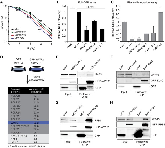
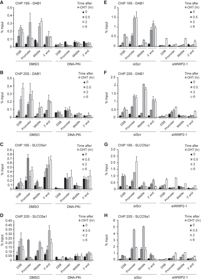
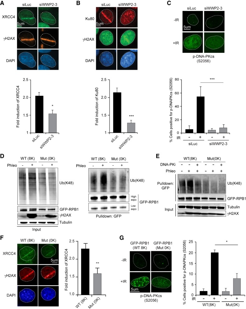
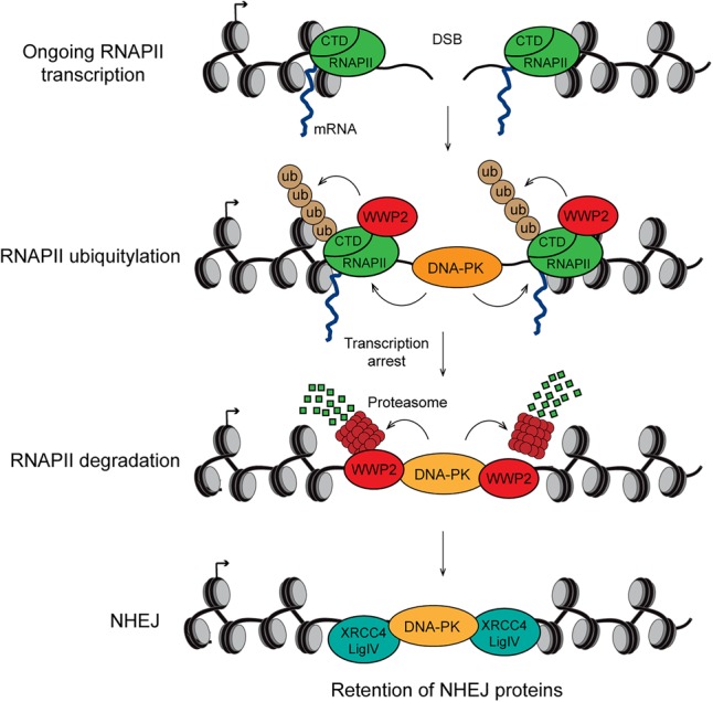
DNA double-strand breaks (DSBs) at RNA polymerase II (RNAPII) transcribed genes lead to inhibition of transcription. The DNA-dependent protein kinase (DNA-PK) complex plays a pivotal role in transcription inhibition at DSBs by stimulating proteasome-dependent eviction of RNAPII at these lesions. How DNA-PK triggers RNAPII eviction to inhibit transcription at DSBs remains unclear. Here we show that the HECT E3 ubiquitin ligase WWP2 associates with components of the DNA-PK and RNAPII complexes and is recruited to DSBs at RNAPII transcribed genes. In response to DSBs, WWP2 targets the RNAPII subunit RPB1 for K48-linked ubiquitylation, thereby driving DNA-PK- and proteasome-dependent eviction of RNAPII. The lack of WWP2 or expression of nonubiquitylatable RPB1 abrogates the binding of nonhomologous end joining (NHEJ) factors, including DNA-PK and XRCC4/DNA ligase IV, and impairs DSB repair. These findings suggest that WWP2 operates in a DNA-PK-dependent shutoff circuitry for RNAPII clearance that promotes DSB repair by protecting the NHEJ machinery from collision with the transcription machinery.

Keywords: DNA double-strand break repair; DNA-PK; RNAPII ubiquitylation; WWP2 HECT E3 ubiquitin ligase; transcription silencing.


Figures

Figure 1.
Figure 1.
WWP2 protects cells against DSBs by promoting NHEJ. (A) Clonogenic survival of VH10-SV40 cells transfected with the indicated siRNAs and exposed to the indicated doses of IR. The mean ± SD from three independent experiments is shown. Statistical significance was calculated using the Student's t-test. (*) P < 0.05; (**) P < 0.01. (B) Quantification of GFP-positive EJ5-GFP HEK293 cells transfected with the indicated siRNAs. DSBs were induced by transfection of an I-SceI expression vector. The transfection efficiency was corrected by cotransfection with an mCherry expression vector. The mean ± SD from two independent experiments is shown. (C) Quantification of plasmid integration efficiencies in U2OS cells transfected with the indicated siRNAs. The mean ± SD from two independent experiments is shown. (D) SILAC (stable isotope labeling by amino acids in culture)-based mass spectrometry analysis of stable U2OS cells expressing GFP (L) or GFP-WWP2 (H). RNAPII complex members are marked in dark gray, whereas NHEJ factors are indicated in light gray. (E) Pull-downs of the indicated GFP fusion proteins in U2OS cells. Blots were probed for Ku80 and GFP. (F) Pull-downs of the indicated GFP fusion proteins in HeLa cells. Blots were probed for WWP2 and GFP. (G) As in E, except that blots were probed for RPB1 and GFP. (H) As in E, except that blots were probed for WWP2 and GFP.
Figure 2.
Figure 2.
WWP2 is recruited to DSBs in transcribed genes to promote DNA repair. (A) Recruitment of mCherry-WWP2 to multiphoton tracks in U2OS cells. GFP-Ku70 was used as a DNA damage marker. (B) Quantification of A. (C) Schematic of the HA-ER-I-PpoI system in U2OS cells used to generate site-specific DSBs at the indicated genes following 4-hydroxytamoxifen (4-OHT) treatment. Gray boxes indicate positions where protein binding is monitored by ChIP-qPCR (chromatin immunoprecipitation [ChIP] combined with quantitative PCR [qPCR]). Black boxes indicate positions of the primers used to quantify mRNA levels of the indicated genes by RT-qPCR. (D) ChIP-qPCR against WWP2 in U2OS HA-ER-I-PpoI cells at the indicated time points after 4-OHT treatment and at the indicated positions at DAB1 and SLCO5a1. The mean ± SD from qPCR replicates of a representative experiment is shown. A repeat of the experiment is shown in Supplemental Figure S2C. (E) Western blot analysis of RPB1 and Ser2- and Ser5-phosphorylated RPB1 (S2 and S5) levels in phleomycin (Phleo)-treated U2OS cells that were left untreated or were treated with 5,6-dichloro-1-β-D-ribofuranosylbenzimidazole (DRB). Tubulin was used as a loading control. (F) Recruitment of GFP-WWP2 to multiphoton tracks in untreated and DRB-treated U2OS cells. (G) Quantification of F. (H) Cutting efficiencies at DAB1 and SLCO5a1 at the indicated time points after 4-OHT treatment in U2OS HA-ER-I-PpoI cells transfected with the indicated siRNAs. The mean ± SD from qPCR replicates of a representative experiment is shown. A repeat of the experiment is shown in Supplemental Figure S8A.
Figure 3.
Figure 3.
WWP2 promotes DSB-induced transcription silencing and RPBI ubiquitylation after DNA damage. (A) RT-qPCR analysis of DAB1 expression levels in U2OS HA-ER-I-PpoI cells at the indicated time points after 4-OHT treatment and transfected with the indicated siRNAs. DAB1 mRNA levels were normalized to those of cyclophilin B. The mean ± SD from qPCR replicates of a representative experiment is shown. A repeat of the experiment is shown in Supplemental Figure S8B. (B) As in A, except for SLCO5a1. A repeat of the experiment is shown in Supplemental Figure S8B. (C) ChIP-qPCR against RPB1 in U2OS HA-ER-I-PpoI cells transfected with the indicated siRNAs. RPB1 levels were monitored at the indicated time points after 4-OHT treatment and at the indicated positions at DAB1. The mean ± SD from qPCR replicates of a representative experiment is shown. A repeat of the experiment is shown in Supplemental Figure S8C. (D) As in C, except for SLCO5a1. A repeat of the experiment is shown in Supplemental Figure S8D. (E) ChIP-qPCR against RPB1 in U2OS HA-ER-I-PpoI cells transfected with the indicated siRNAs. RPB1 levels were monitored at the indicated time points after 4-OHT treatment at the OMA1 and PRKAA2 genes. The mean ± SD from qPCR replicates of a representative experiment is shown. A repeat of the experiment is shown in Supplemental Figure S8E. (F) Pull-downs of GFP-RPB1 under denaturing conditions in untreated and phleomycin (Phleo)-treated U2OS cells. Cells were also treated with proteasome inhibitor (MG-132) 25 min before the phleomycin treatment. Blots were probed for Ub(K48), GFP, and γH2AX. Tubulin was used as a loading control. (G) As in F, except that cells were treated with the indicated siRNAs, and blots were also probed for H3. Relative Ub(K48) levels after GFP-RPB1 pull-down from phleomycin-treated versus untreated cells are indicated below the blots.
Figure 4.
Figure 4.
DNA-PK affects the ubiquitylation and occupancy of RPB1. (A) Pull-downs of GFP-RPB1 under denaturing conditions in phleomycin (Phleo)- and DNA-PK inhibitor (DNA-PKi)-treated U2OS cells. Cells were also treated with proteasome inhibitor (MG-132) 25 min before the phleomycin treatment. Blots were probed for Ub(K48), GFP, H3, and γH2AX. Tubulin was used as a loading control. (B) As in A, except that cells were transfected with the indicated siRNA. (C) ChIP-qPCR against RPB1 and S2-, S5-, or S7-phosphorylated RPB1 (p-RPB1) in DMSO-treated (control) and DNA-PKi-treated U2OS HA-ER-I-PpoI cells at the indicated time points after 4-OHT treatment and at the indicated positions at DAB1. A representative experiment is shown. A repeat of the experiment is shown in Supplemental Figures S10A and S11A. (D) ChIP-qPCR against RPB1 and S2-, S5-, or S7-phosphorylated RPB1 (p-RPB1) in DMSO-treated (control) and DNA-PKi-treated U2OS HA-ER-I-PpoI cells at the indicated time points after 4-OHT treatment and at the indicated positions at SLCO5a1. A representative experiment is shown. A repeat of the experiment is shown in Supplemental Figures S10B and S11B.
Figure 5.
Figure 5.
Proteasomes are recruited to broken genes in a DNA-PKcs- and WWP2-dependent manner. (A) ChIP-qPCR against the 19S proteasome in DMSO-treated (control) and DNA-PKi-treated U2OS HA-ER-I-PpoI cells at the indicated time points after 4-OHT treatment and at the indicated positions at DAB1. The mean ± SD from qPCR replicates of a representative experiment is shown. A repeat of the experiment is shown in Supplemental Figure S13A. (B) As in A, except that the 20S proteasome was examined. A repeat of the experiment is shown in Supplemental Figure S13A. (C) As in A, except for SLCO5a1. A repeat of the experiment is shown in Supplemental Figure S13B. (D) As in B, except for SLCO5a1. A repeat of the experiment is shown in Supplemental Figure S13B. (E) ChIP-qPCR against the 19S proteasome in U2OS HA-ER-I-PpoI cells transfected with the indicated siRNA at the indicated time points after 4-OHT treatment and at the indicated positions at DAB1. The mean ± SD from qPCR replicates of a representative experiment is shown. A repeat of the experiment is shown in Supplemental Figure S14A. (F) As in E, except that the 20S proteasome was examined. A repeat of the experiment is shown in Supplemental Figure S14A. (G) As in E, except for SLCO5a1. A repeat of the experiment is shown in Supplemental Figure S14B. (H) As in F, except for SLCO5a1. A repeat of the experiment is shown in Supplemental Figure S14B.
Figure 6.
Figure 6.
WWP2-dependent RPB1 ubiquitylation promotes accumulation of NHEJ factors at DSBs. (A) Immunofluorescence (IF) images (top panel) and quantification (bottom panel) of XRCC4 recruitment to DNA damage tracks generated by UV-A laser microirradiation in U2OS cells transfected with the indicated siRNAs. γH2AX was used as a DNA damage marker. The mean ± SD from three independent experiments is shown. Statistical significance was calculated using the Student's t-test. (*) P < 0.05. (B) As in A, except for Ku80. The mean ± SD from six independent experiments is shown. Statistical significance was calculated using the Student's t-test. (***) P < 0.001. (C) IF images (top panel) and quantification (bottom panel) of p-DNA-PKcs (S2056) focus formation 1 h after 10 Gy of IR in U2OS cells transfected with the indicated siRNAs. The mean ± S.E.M from four independent experiments is shown. Statistical significance was calculated using the Student's t-test. (***) P < 0.001. (D) Pull-downs of GFP-RPB1 wild type (8K) or mutant (0K) under denaturing conditions in untreated and phleomycin (Phleo)-treated NIH3T3 cells. Cells were also treated with proteasome inhibitor (MG-132) 25 min before the phleomycin treatment. Blots were probed for Ub(K48), GFP, and γH2AX. Tubulin was used as a loading control. (E) As in D, except that cells were also treated with DNA-PKi. (F) IF images (left panel) and quantification (right panel) of XRCC4 recruitment to DNA damage tracks generated by UV-A laser microirradiation in NIH3T3 cells expressing wild-type (8K) or mutant (0K) GFP-RPB1. The mean ± SEM from three independent experiments is shown. Statistical significance was calculated using the Student's t-test. (**) P < 0.01. (G) IF images (left panel) and quantification (right panel) of p-DNA-PKcs (S2056) focus formation 1 h after 10 Gy of IR in NIH3T3 cells expressing wild-type (8K) or mutant (0K) GFP-RPB1. The mean ± SEM from three independent experiments is shown. Statistical significance was calculated using the Student's t-test. (*) P < 0.05.
Figure 7.
Figure 7.
Model of how DNA-PK/WWP2-dependent transcription silencing at DSBs promotes NHEJ. DNA-PK and the HECT E3 ubiquitin ligase WWP2 are recruited to a DSB in a gene that is actively transcribed by RNAPII. DNA-PK effectuates WWP2-dependent K48-linked ubiquitylation of the CTD of RNAPII subunit RPB1 and the subsequent recruitment of the proteasome. The proteasome triggers RNAPII degradation directly on damaged chromatin, thereby silencing transcription of the broken gene. Finally, transcriptional silencing prevents the loss of DNA-PK and downstream NHEJ factors from DSBs, likely by protecting the NHEJ machinery from collision with the transcription machinery, thereby promoting efficient DSB repair via NHEJ.

References

    1. Abu-Zhayia ER, Awwad SW, Ben-Oz BM, Khoury-Haddad H, Ayoub N. 2018. CDYL1 fosters double-strand break-induced transcription silencing and promotes homology-directed repair. J Mol Cell Biol 10: 341–357. 10.1093/jmcb/mjx050 - DOI - PubMed
    1. Agarwal P, Jackson SP. 2016. G9a inhibition potentiates the anti-tumour activity of DNA double-strand break inducing agents by impairing DNA repair independent of p53 status. Cancer Lett 380: 467–475. 10.1016/j.canlet.2016.07.009 - DOI - PMC - PubMed
    1. Awwad SW, Abu-Zhayia ER, Guttmann-Raviv N, Ayoub N. 2017. NELF-E is recruited to DNA double-strand break sites to promote transcriptional repression and repair. EMBO Rep 18: 745–764. 10.15252/embr.201643191 - DOI - PMC - PubMed
    1. Bennardo N, Cheng A, Huang N, Stark JM. 2008. Alternative-NHEJ is a mechanistically distinct pathway of mammalian chromosome break repair. PLoS Genet 4: e1000110 10.1371/journal.pgen.1000110 - DOI - PMC - PubMed
    1. Blackford AN, Jackson SP. 2017. ATM, ATR, and DNA-PK: the trinity at the heart of the DNA damage response. Mol Cell 66: 801–817. 10.1016/j.molcel.2017.05.015 - DOI - PubMed

Publication types

MeSH terms