. 2019 May 2;9(1):6809.
doi: 10.1038/s41598-019-43129-2.

In silico structural elucidation of RNA-dependent RNA polymerase towards the identification of potential Crimean-Congo Hemorrhagic Fever Virus inhibitors

Affiliations

In silico structural elucidation of RNA-dependent RNA polymerase towards the identification of potential Crimean-Congo Hemorrhagic Fever Virus inhibitors

Muhammad Usman Mirza et al. Sci Rep. .

Abstract

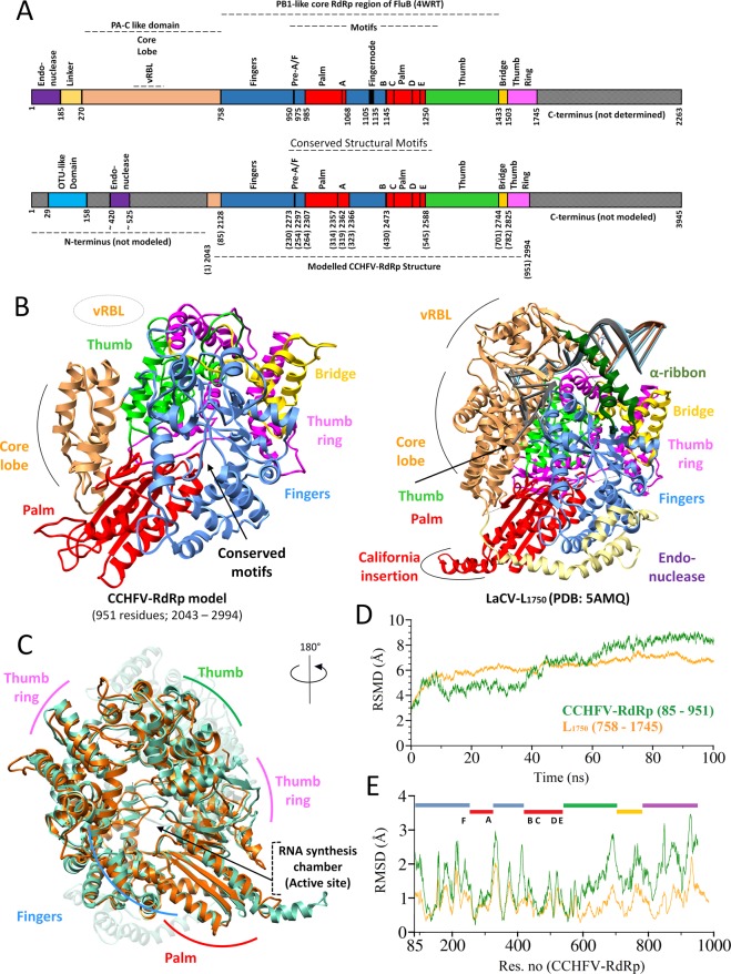
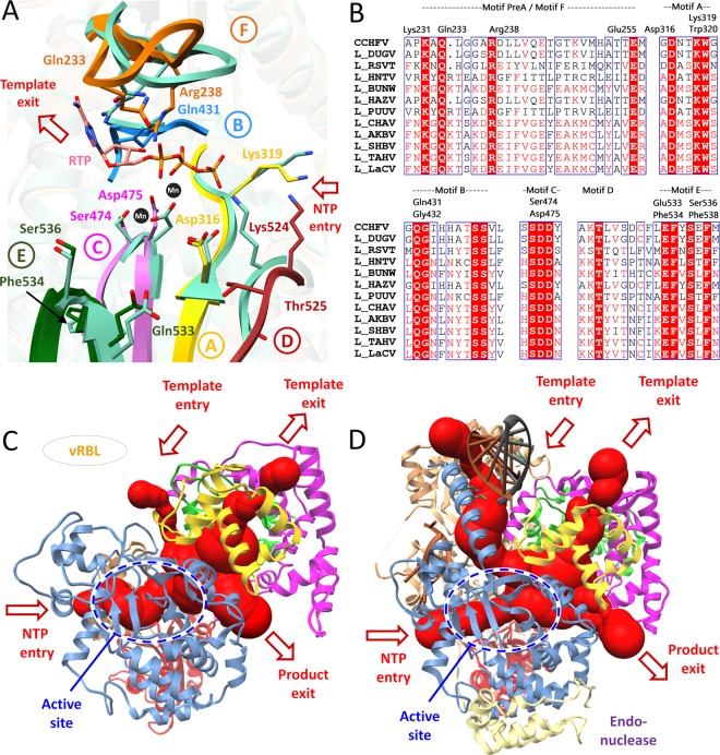
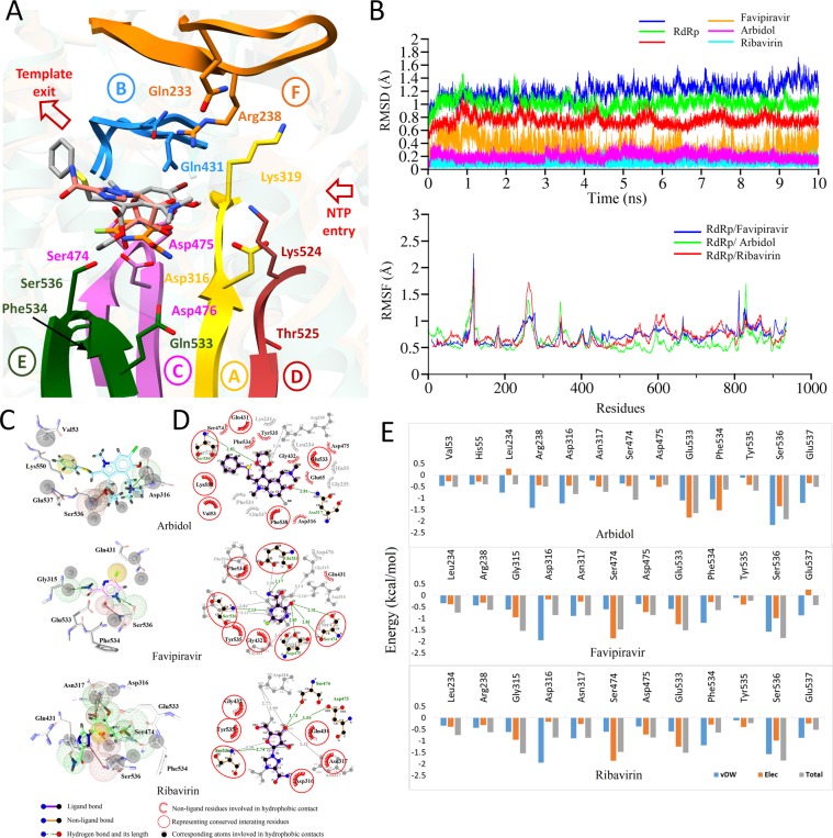
The Crimean-Congo Hemorrhagic Fever virus (CCHFV) is a segmented negative single-stranded RNA virus (-ssRNA) which causes severe hemorrhagic fever in humans with a mortality rate of ~50%. To date, no vaccine has been approved. Treatment is limited to supportive care with few investigational drugs in practice. Previous studies have identified viral RNA dependent RNA Polymerase (RdRp) as a potential drug target due to its significant role in viral replication and transcription. Since no crystal structure is available yet, we report the structural elucidation of CCHFV-RdRp by in-depth homology modeling. Even with low sequence identity, the generated model suggests a similar overall structure as previously reported RdRps. More specifically, the model suggests the presence of structural/functional conserved RdRp motifs for polymerase function, the configuration of uniform spatial arrangement of core RdRp sub-domains, and predicted positively charged entry/exit tunnels, as seen in sNSV polymerases. Extensive pharmacophore modeling based on per-residue energy contribution with investigational drugs allowed the concise mapping of pharmacophoric features and identified potential hits. The combination of pharmacophoric features with interaction energy analysis revealed functionally important residues in the conserved motifs together with in silico predicted common inhibitory binding modes with highly potent reference compounds.


Conflict of interest statement

The authors declare no competing interests.

Figures

Figure 1
Figure 1
Overall homology model of CCHFV-RdRp. (A) Linear schematic representation of domain architecture of CCHFV-RdRp (modelled 2043–2994; 951 residues) (top) aligned to L1750 (bottom), with structurally and functionally conserved domains similarly colored (B) Structural representation of same pose of CCHFV-RdRp model (left) with L1750 (right), elaborating the similarly colored structural homologous features including canonical fingers, palm and thumb subdomains along with other domains (C) Structural superimposition of CCHFV-RdRp (orange) and L1750 (green) highlighting the similar configuration of thumb and fingers subdomains by their positions in space relative to the palm subdomain. The conformation is rotated to display the conserved RNA synthesis chamber (active site). (D) Root Mean Square Deviation (RMSD) of CCHFV-RdRp model (green) in Angstrom (Å) during 100 ns simulations together with the reference L1750 structure. (E) Root Mean Square Fluctuation (RMSF) of all residues of CCHFV-RdRp model along with the reference L1750 structure during MD simulation and represented domains are highlighted with their respective colors (as in (A)).
Figure 2
Figure 2
The predicted CCHFV-RdRp active site and positively charged tunnels. (A) The arrangement of structurally conserved RdRp motifs in CCHFV-RdRp model colored yellow, dogged blue, orchid, brown, green and orange for motifs A-F respectively, and superimposed on L1750 (sea green). (B) The corresponding predicted binding site residues are aligned through multiple structure alignment in corresponding motifs. Superposition of the polio virus elongation complex structure (PDB: 3OL8) and foot-and-mouth disease virus in complex with RTP (PDB: 2E9R) shows the positions of the catalytic divalent cations (black spheres) and RTP (salmon) (C) The CCHFV-RdRp model is shown (in ribbon) with four predicted positively charged tunnels (red) marked with arrows as entrance (template and NTP entry channel) and exit (template and nascent strand exit channel) tunnels calculated with MOLE2.0. The domains are same as colored in Fig. 1A. (D) The same representation as (C) for L1750 (PDB: 5 amq) with the 5′ and 3′ vRNA is highlighted. Domain colors are same as in Fig. 1A.
Figure 3
Figure 3
Per-residue energy distribution-based pharmacophore model. (A) MD simulated conformations of ribavirin (salmon), arbidol (grey) and favipiravir (orange) in sticks, inside the predicted binding pocket of CCHFV-RdRp. The arrangement of motifs and colors are same as in Fig. 2A. (B) Root Mean Square Deviation (RMSD) of CCHFV-RdRp complexed with all three reported drugs with distinctive colors are highlighted over a period of 10 ns simulations, while, Root Mean Square Fluctuation (RMSF) of all residues during MD simulation are highlighted below. (C) Pharmacophoric features projected within the predicted target site of bound ligand with H-bond donors (HBDs) and H-bond acceptors (HBAs) are highlighted in green and red spheres, respectively. Hydrophobic centers are in yellow and exclusion volume spheres are highlighted in grey. (D) 2D interaction plot for all three reported drugs complexed with RdRp. (E) Per-residue decomposition analysis performed with Amber 16 are presented in bar chart for highly contributing residues of predicted binding site of CCHFV-RdRp.
Figure 4
Figure 4
Post-Molecular dynamics (MD) analysis of CCHFV-RdRp complexes. (A) MD simulated conformations of cmd1, cmd2, cmd3, and two reference compounds, RTP and 2′FdC in the sticks, inside the predicted binding pocket of CCHFV-RdRp. The arrangement of motifs and colors are same as in Fig. 3A. (B) Root Mean Square Deviation (RMSD) of CCHFV-RdRp complexed with presented compounds with distinctive colors are highlighted over a period of 10 ns simulations, while, Root Mean Square Fluctuation (RMSF) of all residues during MD simulation are highlighted below. (C) 2D interaction plot is interactively displayed for all presented compounds complexed with RdRp. (D) Per-residue decomposition analysis for all 5 compounds performed with Amber 16 is presented in a bar chart for highly contributing residues of the predicted binding site of CCHFV-RdRp. (E) H-bond occupancy of particular amino acids in frames throughout 10 ns is displayed for cmd1/2 and 3.

References

    1. Dokuzoguz B, et al. Severity scoring index for Crimean-Congo hemorrhagic fever and the impact of ribavirin and corticosteroids on fatality. Clinical infectious diseases. 2013;57:1270–1274. doi: 10.1093/cid/cit527. - DOI - PubMed
    1. Ergönül Ö. Crimean-Congo haemorrhagic fever. The Lancet Infectious Diseases. 2006;6:203–214. doi: 10.1016/s1473-3099(06)70435-2. - DOI - PMC - PubMed
    1. Deyde VM, Khristova ML, Rollin PE, Ksiazek TG, Nichol ST. Crimean-Congo hemorrhagic fever virus genomics and global diversity. J Virol. 2006;80:8834–8842. doi: 10.1128/JVI.00752-06. - DOI - PMC - PubMed
    1. Adams MJ, et al. Changes to taxonomy and the International Code of Virus Classification and Nomenclature ratified by the International Committee on Taxonomy of Viruses (2017) Archives of Virology. 2017;162:2505–2538. doi: 10.1007/s00705-017-3358-5. - DOI - PubMed
    1. Burt FJ, Spencer DC, Leman PA, Patterson B, Swanepoel R. Investigation of tick-borne viruses as pathogens of humans in South Africa and evidence of Dugbe virus infection in a patient with prolonged thrombocytopenia. Epidemiology and Infection. 1996;116:353–361. doi: 10.1017/S0950268800052687. - DOI - PMC - PubMed

Publication types

MeSH terms