. 2019 Mar 27:2019:7947596.
doi: 10.1155/2019/7947596. eCollection 2019.

Elevation of IL-6 and IL-33 Levels in Serum Associated with Lung Fibrosis and Skeletal Muscle Wasting in a Bleomycin-Induced Lung Injury Mouse Model

Affiliations

Elevation of IL-6 and IL-33 Levels in Serum Associated with Lung Fibrosis and Skeletal Muscle Wasting in a Bleomycin-Induced Lung Injury Mouse Model

Jiunn-Min Shieh et al. Mediators Inflamm. .

Abstract

Weight loss due to skeletal muscle atrophy in patients with chronic pulmonary disease is negatively correlated with clinical outcome. Pulmonary fibrosis is a chronic and progressive interstitial lung disease characterized by the dysregulated deposition of the extracellular matrix (ECM) with the destruction of normal tissue, resulting in end-stage organ failure. BLM-induced fibrosis is one of several different experimental models of pulmonary fibrosis, characterized by inflammation and excessive ECM deposition. We directly induced mouse lung injury by the intratracheal administration of bleomycin and monitored the physiological and biochemical changes in lung and skeletal muscle tissues by using lung function testing, ELISA, Western blotting, and immunohistochemistry. Here, we found that BLM-induced lung fibrosis with thickened interstitial lung tissue, including fibronectin and collagen, was correlated with the increased serum concentrations of IL-6 and IL-33 and accompanied by reduced lung function, including FRC (functional residual capacity), C chord (lung compliance), IC (inspiratory capacity), VC (vital capacity), TLC (total lung capacity), and FVC (forced vital capacity) (p < 0.05). The activity of AKT in lung tissue was suppressed, but conversely, the activity of STAT3 was enhanced during lung fibrosis in mice. In addition, we found that the amount of sST2, the soluble form of the IL-33 receptor, was dramatically decreased in lung fibrosis tissues. The skeletal muscle tissue isolated from lung injury mice increased the activation of STAT3 and AMPK, accompanied by an increased amount of Atrogin-1 protein in BLM-induced lung fibrosis mice. The mouse myoblast cell-based model showed that IL-6 and IL-33 specifically activated STAT3 and AMPK signaling, respectively, to induce the expression of the muscle-specific proteolysis markers MuRF1 and Atrogin-1. These data suggested that increased levels of IL-6 and IL-33 in the serum of mice with BLM-induced lung injury may cause lung fibrosis with thickened interstitial lung tissue accompanied by reduced lung function and muscle mass through the activation of STAT3 and AMPK signals.


Figures

Figure 1
Figure 1
BLM-induced lung fibrosis in mice. C57BL/6 mice were intratracheally administered bleomycin (2 mg/kg) for 14 days. (a–d) Lung H&E staining. (e–h) Masson's trichrome staining of lung tissue from (a, c) control mice and (b, d) BLM-induced lung fibrosis mice. The photos in (a, b, e, and f) represent 50x magnification. The photos in (c, d, g, and h) represent 200x magnification. The collagen in lung fibrosis tissue was stained blue by Masson's trichrome staining. Bar = 200 μm at 50x magnification and 50 μm at 200x magnification as indicated.
Figure 2
Figure 2
Lung function was reduced in mice with BLM-induced fibrotic lungs. Anesthetized mice were endotracheally intubated with an airway catheter, and the parameters of lung function were measured by a pulmonary function testing system. (a) FVC (forced vital capacity), (b) IC (inspiratory capacity), (c) VC (vital capacity), (d) FRC (functional residual capacity), (e) TLC (total lung capacity), and (f) C chord (lung compliance). The quantitative data were expressed as the means ± SD. Light spots represent the lung function of control mice (n = 4), and dark spots represent the lung function of mice with BLM-induced lung injury (n = 5). p < 0.05 and ∗∗p < 0.005 compared with control mice.
Figure 3
Figure 3
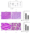
BLM-induced lung injury may cause body weight loss and muscle atrophy. (a) The body weights of experimental mice were measured every two to three days during the experimental periods. Square boxes represent the body weight change of control mice (n = 6), and dots represent the body weight change of mice with BLM-induced lung injury (n = 5). The quantitative data were expressed as the means ± SD. p < 0.05 compared with control. (b–g) Cross-sectional H&E staining of quadriceps muscle in (b, e) control mice and (c, f) mice with BLM-induced lung fibrosis. The photos represent 50x magnification in (b–d) and 200x magnification in (e–g). The quantitative analysis of the muscle bundle area to the total area in photos is shown in (d, g) (n = 3). The values of all data are averages from at least three photos with different fields of view. The muscle bundle area was measured by ImageJ software. p < 0.05 compared with control mice. Bar = 200 μm at 50x magnification and 50 μm at 200x magnification as indicated. The black arrow indicates the detached basal lamina.
Figure 4
Figure 4
The circulation of IL-6 and IL-33 was increased in mice with BLM-induced lung fibrosis. (a) Protein levels in bronchoalveolar lavage fluid (BALF) were estimated by using a BCA kit assay. The cytokines in mouse serum were measured by ELISA. (b) IL-6. (c) IL-33. Light spots represent the cytokine amount in control mice (n = 4), and dark spots represent the cytokine amount in mice with BLM-induced lung injury (n = 5). The quantitative data were expressed as the means ± SD. p < 0.05 compared with control.
Figure 5
Figure 5
Activation of STAT3 is associated with inactivation of AKT in fibrotic lung tissue. The sliced left lung lobes of mice were homogenized, and the lysate was analyzed by Western blotting. (a) Fibronectin and STAT3. (b) ST2and AKT were detected in lung lysates. The intensity of bands in the Western blots was measured by ImageJ software. GAPDH was used as an internal control. The quantitative data were expressed as the means ± SD. p < 0.05 compared with control.
Figure 6
Figure 6
IL-6 and IL-33 may synergistically cause muscle atrophy. (a) The quadriceps muscle of the mice was homogenized, and the lysate was analyzed by Western blotting with specific antibodies against STAT3, AMPK, and Atrogin-1. (b) C2C12 cells, mouse adherent myoblasts, were incubated with 2% horse serum for 72 hours and stimulated with recombinant mouse IL-6 and IL-33 in serum-free medium as indicated for 24 hours. The remaining cells were harvested, and the levels of p-STAT3, STAT3, p-AMPKα, and AMPKα in the cell lysate were analyzed by Western blotting. α-Tubulin and GAPDH were used as internal controls. The intensity of bands in the Western blots was measured by ImageJ software. The quantitative data were expressed as the means ± SD. p < 0.05 compared with control.
Figure 7
Figure 7
Increased levels of IL-6 and IL-33 in the serum of mice with BLM-induced lung injury may cause body weight loss through the activation of STAT3 and AMPK signals. Schematic representation of the role of IL-6 and IL-33 in the BLM-induced lung fibrosis mouse model. IL-6 and IL-33 are secreted from inflammatory lung tissue and modulate lung remodeling, including myofibroblast proliferation, ECM deposition, and decreased lung function. In addition, IL-6 and IL-33 synergistically cause body weight loss in mice through the activation of STAT3 and AMPK signaling to induce the expression of proteolysis- and lipolysis-related genes.

References

    1. Raghu G., Collard H. R., Egan J. J., et al. An official ATS/ERS/JRS/ALAT statement: idiopathic pulmonary fibrosis: evidence-based guidelines for diagnosis and management. American Journal of Respiratory and Critical Care Medicine. 2011;183(6):788–824. doi: 10.1164/rccm.2009-040GL. - DOI - PMC - PubMed
    1. King T. E., Jr., Pardo A., Selman M. Idiopathic pulmonary fibrosis. The Lancet. 2011;378(9807):1949–1961. doi: 10.1016/S0140-6736(11)60052-4. - DOI - PubMed
    1. Richeldi L., HRHR D., Spagnolo P., Luppi F., Cochrane Airways Group Corticosteroids for idiopathic pulmonary fibrosis. Cochrane Database of Systematic Reviews. 2003;3 doi: 10.1002/14651858.CD002880. - DOI - PMC - PubMed
    1. Raghu G., Depaso W. J., Cain K., et al. Azathioprine combined with prednisone in the treatment of idiopathic pulmonary fibrosis: a prospective double-blind, randomized, placebo-controlled clinical trial. American Review of Respiratory Disease. 1991;144(2):291–296. doi: 10.1164/ajrccm/144.2.291. - DOI - PubMed
    1. Collard H. R., Ryu J. H., Douglas W. W., et al. Combined corticosteroid and cyclophosphamide therapy does not alter survival in idiopathic pulmonary fibrosis. Chest. 2004;125(6):2169–2174. doi: 10.1378/chest.125.6.2169. - DOI - PubMed

LinkOut - more resources