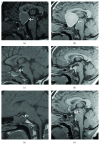
Causes and Follow-Up of Central Diabetes Insipidus in Children
- PMID: 31049061
- PMCID: PMC6458924
- DOI: 10.1155/2019/5303765
Causes and Follow-Up of Central Diabetes Insipidus in Children
Abstract
Objective: To identify the causes of central diabetes insipidus (CDI) by evaluating the values of magnetic resonance imaging (MRI) in the diagnosis of pediatric CDI, providing evidence for the clinical diagnosis and treatment of CDI.
Methods: Seventy-nine patients with CDI (CDI group) hospitalized from July 2012 to March 2017 and 43 healthy children (control group) were enrolled in this study. All cases underwent MRI examination including T1-weighted three-dimensional magnetization-prepared rapid gradient-echo (T1WI-3D-MP RAGE) imaging sequences. The pituitary volume, the signal intensity of posterior pituitary, and the morphology of pituitary stalk were measured between two groups. The medical history, urine testing, imaging of hypothalamic-pituitary region, and hormone levels were also recorded.
Results: Age and gender were matched between the CDI and control groups. The height and BMI in the CDI group were less and the urine volume in 24 h was higher than those in the control group. The signal intensity of the posterior pituitary was higher in the control group, whereas the pituitary volume was smaller in the CDI group. In the CDI group, 44 cases presented with morphological changes of the pituitary stalk. Clinical symptoms mainly included polydipsia, polyuria, short stature, and vomiting. All patients were confirmed by water deprivation vasopressin test. Forty-four CDI children were associated with hypopituitarism, including 33 cases of PSIS with multiple pituitary hormone deficiencies (MPHD) and 11 cases of growth hormone deficiency (IGHD). The pituitary volume in the cases of pituitary stalk interruption syndrome (PSIS) with MPHD was smaller than that in the IGHD patients.
Conclusions: The signal intensity ratio of the posterior lobe, pituitary volume, and the morphology of pituitary stalk on T1WI-3D-MP RAGE image contribute to the diagnosis of CDI.
Figures


Similar articles
-
Clinico-radiological correlation of pituitary stalk interruption syndrome in children with growth hormone deficiency.Pituitary. 2023 Oct;26(5):622-628. doi: 10.1007/s11102-023-01351-2. Epub 2023 Sep 11. Pituitary. 2023. PMID: 37695468
-
Pituitary Morphology and Function in 43 Children with Central Diabetes Insipidus.Int J Endocrinol. 2016;2016:6365830. doi: 10.1155/2016/6365830. Epub 2016 Mar 29. Int J Endocrinol. 2016. PMID: 27118970 Free PMC article.
-
Approach to the Pediatric Patient: Central Diabetes Insipidus.J Clin Endocrinol Metab. 2022 Apr 19;107(5):1407-1416. doi: 10.1210/clinem/dgab930. J Clin Endocrinol Metab. 2022. PMID: 34993537
-
Neuroimaging of central diabetes insipidus.Handb Clin Neurol. 2021;181:207-237. doi: 10.1016/B978-0-12-820683-6.00016-6. Handb Clin Neurol. 2021. PMID: 34238459 Review.
-
Central diabetes insipidus in children: Diagnosis and management.Best Pract Res Clin Endocrinol Metab. 2020 Sep;34(5):101440. doi: 10.1016/j.beem.2020.101440. Epub 2020 Jun 29. Best Pract Res Clin Endocrinol Metab. 2020. PMID: 32646670 Review.
Cited by
-
Central diabetes insipidus developing in a 6-year-old patient 4 years after the remission of unifocal bone Langerhans cell histiocytosis.Clin Pediatr Endocrinol. 2021;30(3):149-153. doi: 10.1297/cpe.30.149. Epub 2021 Jul 10. Clin Pediatr Endocrinol. 2021. PMID: 34285458 Free PMC article.
-
Diabetes Insipidus: A Pragmatic Approach to Management.Cureus. 2021 Jan 5;13(1):e12498. doi: 10.7759/cureus.12498. Cureus. 2021. PMID: 33425560 Free PMC article. Review.
-
Clinical Characteristics of Pediatric Patients With Sellar and Suprasellar Lesions Who Initially Present With Central Diabetes Insipidus: A Retrospective Study of 55 Cases From a Large Pituitary Center in China.Front Endocrinol (Lausanne). 2020 Feb 20;11:76. doi: 10.3389/fendo.2020.00076. eCollection 2020. Front Endocrinol (Lausanne). 2020. PMID: 32153511 Free PMC article.
-
Incidence and Predictors for Oncologic Etiologies in Chinese Children with Pituitary Stalk Thickening.Cancers (Basel). 2023 Aug 2;15(15):3935. doi: 10.3390/cancers15153935. Cancers (Basel). 2023. PMID: 37568752 Free PMC article.
-
Deficiency of arginine vasopressin in children - diagnostic and therapeutic approaches to improve patients' quality of life based on a 25-year, single-centre retrospective analysis.Pediatr Endocrinol Diabetes Metab. 2024;30(4):198-210. doi: 10.5114/pedm.2024.146684. Pediatr Endocrinol Diabetes Metab. 2024. PMID: 39963057 Free PMC article.
References
-
- Moritz M. L., Ayus J. C. Textbook of Nephro-Endocrinology. Academic Press; 2018. Diabetes insipidus and syndrome of inappropriate antidiuretic hormone; pp. 133–161. - DOI
-
- León-Suárez A., Roldán-Sarmiento P., Gómez-Sámano M. A., et al. Infundibulo-hypophysitis-like radiological image in a patient with pituitary infiltration of a diffuse large B-cell non-Hodgkin lymphoma. Endocrinology, Diabetes & Metabolism Case Reports. 2016;2016:p. 2016. doi: 10.1530/edm-16-0103. - DOI - PMC - PubMed
-
- Lazzari I., Graziani A., Mirici F., Stefanini G. F. Transient idiopathic central diabetes insipidus: is severe sepsis a possible cause? Italian Journal of Medicine. 2017;11(1):p. 78. doi: 10.4081/itjm.2017.717. - DOI
LinkOut - more resources
Full Text Sources

