. 2019 Mar 27:2019:8034578.
doi: 10.1155/2019/8034578. eCollection 2019.

In Vitro Effect of Estradiol and Progesterone on Ovine Amniotic Epithelial Cells

Affiliations

In Vitro Effect of Estradiol and Progesterone on Ovine Amniotic Epithelial Cells

Annunziata Mauro et al. Stem Cells Int. .

Abstract

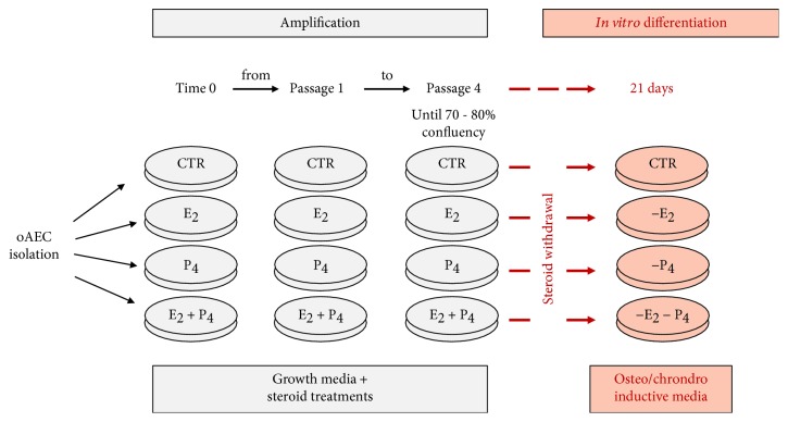
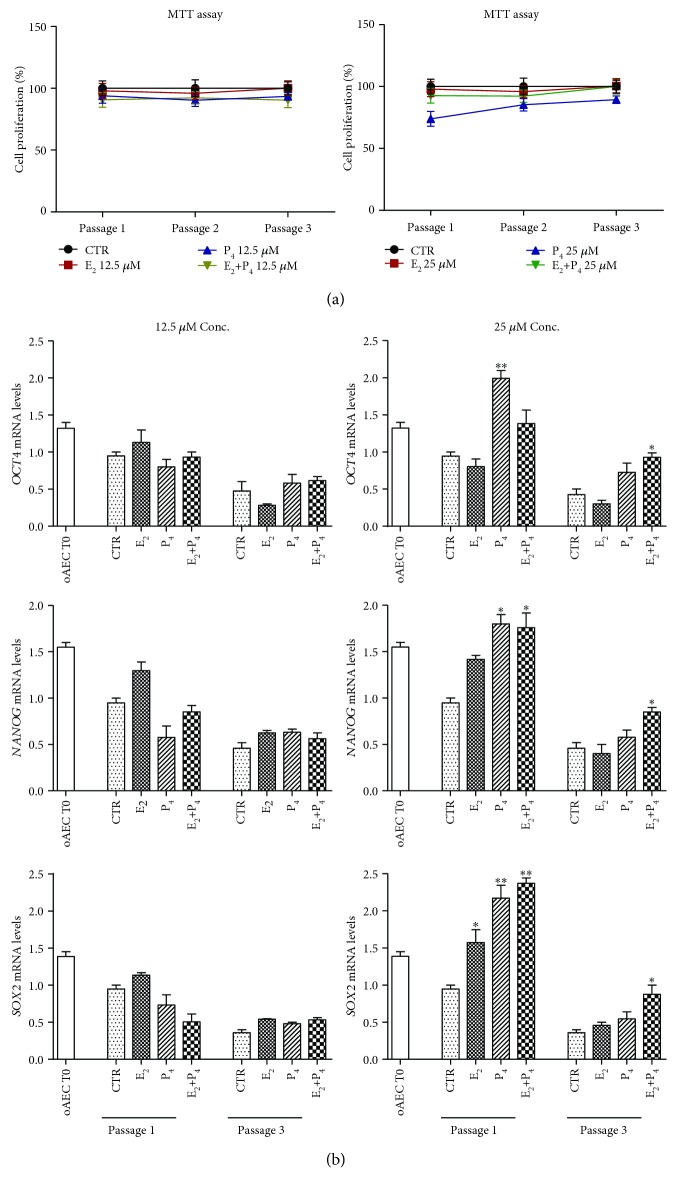
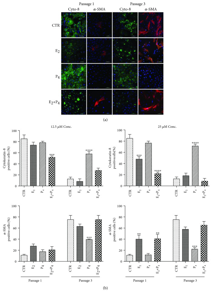
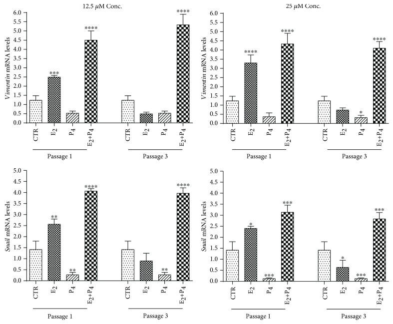
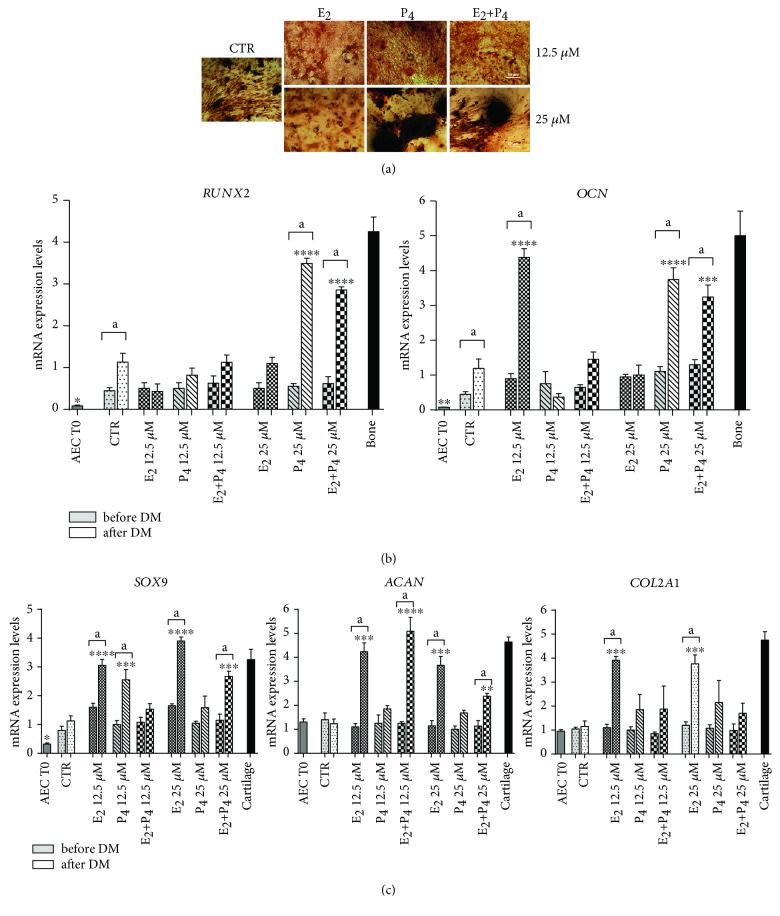
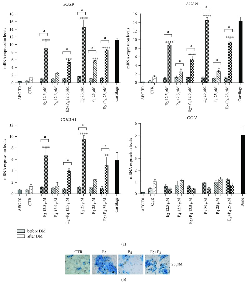
Amniotic epithelial cells (AECs), an emerging source of extrafoetal stem cells, have recently attracted attention for their great regenerative potential. Since AEC amplifications are accompanied by the loss of their native epithelial phenotype and by the progressive reduction of relevant biological properties, the issue to be addressed is the development of effective culture protocols. In this context, recently, it has been demonstrated that progesterone (P4) supplementation during ovine AEC (oAEC) expansion could prevent the undesirable epithelial-mesenchymal transition (EMT). In contrast, there is no information to date on the role of the other pregnancy steroids in culture. With this aim, the present study has been designed to clarify the impact of estradiol (E2), alone or in combination with P4 (12.5 μM and 25 μM), during oAEC amplification. Steroid supplementations were assessed by testing oAEC proliferation, stemness, EMT, and osteogenic or chondrogenic plasticity. The results indicated that EMT can be prevented exclusively in the presence of high doses of P4, while it occurred rapidly in cells exposed to E2 as denoted by protein (cytokeratin-8 and alpha-SMA) and gene expression (vimentin and snail) profiles. Moreover, steroid exposure was able to influence highly oAEC plasticity. Particularly, P4-treated cells displayed a precommitment towards osteogenic lineage, confirmed by the upregulation of OCN, RUNX2, and the greater deposition of calcium nodules. Conversely, P4 exposure inhibited oAEC chondrogenic differentiation, which was induced in E2-treated cells as confirmed by the upregulation of chondrogenesis-related genes (SOX9, ACAN, and COL2A1) and by the accumulation of Alcian blue-positive extracellular matrix. Simultaneously, E2-treated cells remained unresponsive to osteogenic inductive stimuli. In conclusion, media supplementation with high doses of steroids may be adopted to modulate phenotype and plasticity during oAEC amplification. Relevantly, the osteo or chondro steroid-induced precommitment may open unprecedented cell-based therapies to face the unsolved orthopaedic issues related to osteochondral regeneration.


Figures

Figure 1
Figure 1
Schematic representation of the experimental design. Under the grey banner, the amplification protocols are briefly summarized: starting from freshly isolated oAEC, cells were cultured in growth medium in the absence (CTR) or in presence of different steroid treatments: E2 and P4 alone or in combination (E2+P4) at two concentrations (12.5 μM or 25 μM). This incubation, aimed at promoting cell expansion, was carried out until passage 4. Under the pink banner is the summarized differentiation procedures: at passage 4, once oAEC reached 70-80% of confluency, steroids were withdrawn and the cells were incubated for 21 days under differentiation conditions by exposing them to osteo- or chondrodifferentiation medium. oAECs: ovine amniotic epithelial cells; CTR: control cell; E2: estradiol; P4: progesterone.
Figure 2
Figure 2
Effect of steroids on doubling time and stemness gene expression in oAECs. (a) Proliferation activity of CTR and steroid-treated oAEC (E2, P4, and E2+P4) at 12.5 μM or 25 μM during in vitro amplification. The data are expressed as percentage of proliferation ± SEM from values of triplicate samples obtained by three different animals, CTR set to 100%. (b) Real-time qPCR analysis of stemness gene expression profile (OCT4, SOX2, and NANOG) in CTR and steroid-treated oAEC at passage 1 and passage 3. Freshly isolated oAECs (AEC T0) were used as the internal control of stemness gene expression. Relative quantification of each mRNA gene expression was calculated using the ΔΔCt method and presented as fold change in gene expression normalized to endogenous GAPDH (internal control) and relative to the CTR at passage 1 (calibrator). Data was expressed as mean ± SEM values of samples, each performed in triplicate, obtained at least three different animals. Values were considered statistically significant for p < 0.05 and ∗∗p < 0.01 with respect to the CTR values within the same passage of culture. oAECs: ovine amniotic epithelial cells; T0: time zero; CTR: control cell; E2: estradiol; P4: progesterone.
Figure 3
Figure 3
EMT steroid modulation on amplified oAEC. (a) Immunostaining for cytokeratin-8 (green) and α-SMA (red), epithelial and mesenchyme markers, respectively, was performed on CTR and steroid-amplified oAEC (E2, P4, and E2+P4 at 25 μM), thus demonstrating that both protein profiles changed in a steroid- and passage-dependent manner (passage 1 and passage 3). Nuclei are counterstained with DAPI (blue). Scale bar: 50 μm. (b) Fluorescence quantification of cytokeratin-8- and α-SMA-positive cells recorded in CTR and steroid-treated oAEC. This analysis was performed on cells amplified with different combinations and concentrations (12.5 μM and 25 μM) of steroids from passage 1 to passage 3. The data are expressed as mean ± SEM values of samples, performed in triplicate, obtained at least three different animals. Values statistically different for p < 0.05, ∗∗p < 0.01, and ∗∗∗p < 0.001 in comparison to CTR within each passage. CTR: control cell; E2: estradiol; P4: progesterone; Cyto-8: cytokeratin-8; α-SMA: alpha-smooth muscle actin.
Figure 4
Figure 4
Influence of steroids on EMT gene expression during oAEC amplification. Real-time qPCR analysis of two EMT-related gene expressions (vimentin and snail) was carried out on oAEC amplified under CTR conditions or exposed to steroids at 12.5 μM or 25 μM (E2, P4, and E2+P4) up to passage 3. Relative quantification of each mRNA gene expression was calculated using the ΔΔCt method and presented as fold change in gene expression normalized to endogenous GAPDH (internal control) and relative to the CTR at passage 1 (calibrator). Data was expressed as mean ± SEM values of samples, each performed in triplicate, obtained at least three different animals. Values statistically different for p < 0.05, ∗∗p < 0.01, ∗∗∗p < 0.001, and ∗∗∗p < 0.0001. CTR: control cell; E2: estradiol; P4: progesterone.
Figure 5
Figure 5
Response of steroids amplified oAEC to osteogenic differentiation. (a) Alizarin red staining was used to evaluate deposition of mineralized matrix nodules in CTR and steroid pretreated oAEC (E2, P4, or E2+P4 treatments at 25 μM) after 21 days of culture in osteogenic media (DM). Scale bar = 50 μm. (b) Expression of bone-related genes (RUNX2 and OCN) analysed by real-time qPCR analysis in oAEC after isolation (time 0) and in CTR and steroid pretreated oAEC before (before DM) and after (after DM) osteogenic differentiation. Ovine bone tissue was used as positive control for bone-related genes. (c) Expression of an early (SOX9) and two late chondrogenesis-related genes (ACAN and COL2A1) by real-time qPCR analysis in oAEC after isolation (time 0) and in CTR and steroid pretreated oAEC before (before DM) and after (after DM) osteogenic differentiation. Cartilage tissue was used as positive control for chondrogenesis-related genes. Relative quantification of each mRNA gene expression was calculated using the ΔΔCt method and presented as fold change in gene expression normalized to endogenous GAPDH (internal control) and relative to the CTR after DM (calibrator). Data was expressed as mean ± SEM values of samples, each performed in triplicate, obtained at least three different animals. Values statistically different for p < 0.05, ∗∗p < 0.01, ∗∗∗p < 0.001, and ∗∗∗p < 0.0001 vs. CTR after DM. aValues statistically different for p < 0.05 in the same sample before and after DM. CTR: control cell; E2: estradiol; P4: progesterone.
Figure 6
Figure 6
Response of steroids amplified oAEC to chondrogenic inductive cultural conditions. (a) Real-time qPCR analysis of SOX9, COL2A1, ACAN, and OCN expression in freshly isolated oAEC (time 0) and in differentiated CTR and steroid pretreated oAEC (E2, P4, or E2+P4 at 12.5 μM and 25 μM) after in vitro culture in chondrogenic medium for 21 days. Cartilage and bone tissues were used as positive control for gene expression. Relative quantification of each mRNA gene expression was calculated using the ΔΔCt method and presented as fold change in gene expression normalized to endogenous GAPDH (internal control) and relative to the CTR after DM (calibrator). Data was expressed as mean ± SEM values of samples, each performed in triplicate, obtained at least three different animals. Values statistically different for p < 0.05, ∗∗p < 0.01, ∗∗∗p < 0.001, and ∗∗∗p < 0.0001 vs. CTR after DM. aValues statistically different for p < 0.05 in the same sample before and after DM. (b) Representative images of Alcian blue staining to assess deposition of proteoglycans in the extracellular matrix in CTR and steroid pretreated oAEC (E2, P4, or E2+P4 at 25 μM) after chondrogenic differentiation. Scale bar = 50 μm. CTR: control cell; E2: estradiol; P4: progesterone.

Similar articles

Cited by

References

    1. Parolini O., Soncini M., Evangelista M., Schmidt D. Amniotic membrane and amniotic fluid-derived cells: potential tools for regenerative medicine? Regenerative Medicine. 2009;4(2):275–291. doi: 10.2217/17460751.4.2.275. - DOI - PubMed
    1. Malek A., Bersinger N. A. Human placental stem cells: biomedical potential and clinical relevance. Journal of Stem Cells. 2011;6(2):75–92. - PubMed
    1. Miki T. Amnion-derived stem cells: in quest of clinical applications. Stem Cell Research & Therapy. 2011;2(3):p. 25. doi: 10.1186/scrt66. - DOI - PMC - PubMed
    1. Barboni B., Curini V., Russo V., et al. Indirect co-culture with tendons or tenocytes can program amniotic epithelial cells towards stepwise tenogenic differentiation. PLoS One. 2012;7(2, article e30974) doi: 10.1371/journal.pone.0030974. - DOI - PMC - PubMed
    1. Mamede A. C., Carvalho M. J., Abrantes A. M., Laranjo M., Maia C. J., Botelho M. F. Amniotic membrane: from structure and functions to clinical applications. Cell and Tissue Research. 2012;349(2):447–458. doi: 10.1007/s00441-012-1424-6. - DOI - PubMed