. 2019 Apr 22:11:348-354.
doi: 10.1016/j.dadm.2019.03.002. eCollection 2019 Dec.

Nonparenchymal fluid is the source of increased mean diffusivity in preclinical Alzheimer's disease

Affiliations

Nonparenchymal fluid is the source of increased mean diffusivity in preclinical Alzheimer's disease

Farshid Sepehrband et al. Alzheimers Dement (Amst). .

Abstract

Introduction: Although increased mean diffusivity of the white matter has been repeatedly linked to Alzheimer's disease pathology, the underlying mechanism is not known.

Methods: Here, we used ADNI-3 multishell diffusion magnetic resonance imaging data to separate the diffusion signal of the parenchyma from less hindered fluid pools within the white matter such as perivascular space fluid and fluid-filled cavities.

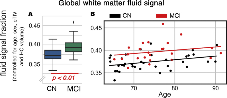
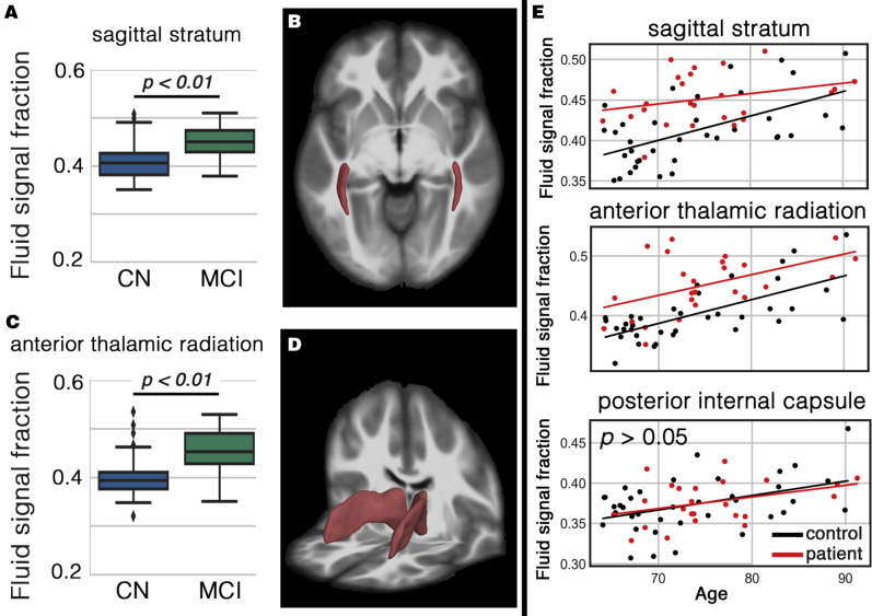
Results: We found that the source of the pathological increase of the mean diffusivity is the increased nonparenchymal fluid, often found in lacunes and perivascular spaces. In this cohort, the cognitive decline was significantly associated with the fluid increase and not with the microstructural changes of the white matter parenchyma itself. The white matter fluid increase was dominantly observed in the sagittal stratum and anterior thalamic radiation.

Discussion: These findings are positive steps toward understanding the pathophysiology of white matter alteration and its role in the cognitive decline.

Keywords: Nonparenchymal fluid; Preclinical AD; White matter alteration; White matter fluid.


Figures

Fig. 1
Fig. 1
Global white matter fluid signal (WFS) difference between cognitively normal (CN) and mild cognitively impaired (MCI) groups. (A) Mean global difference between CN and MCI, controlled for age, sex, eTIV, and the hippocampal (HC) volume. (B) The WFS changes as functions of cognitive state and age are demonstrated.
Fig. 2
Fig. 2
White matter regions with significant higher white matter fluid signal (WFS) in cognitively impaired subjects. A shows the increased WFS in sagittal stratum. Sagittal stratum area is illustrated in B. Scatter plots C and D are related to the anterior thalamic area. Last column (E) shows the WFS of both groups as a function of age. Posterior internal capsule included as an example with no statistical difference to appreciate the extent of change in the affected white matter regions.

References

    1. Brun A., Englund E. A white matter disorder in dementia of the Alzheimer type: A pathoanatomical study. Ann Neurol. 1986;19:253–262. - PubMed
    1. Zlokovic B.V. Neurovascular pathways to neurodegeneration in Alzheimer's disease and other disorders. Nat Rev Neurosci. 2011;12:723–738. - PMC - PubMed
    1. Iturria-Medina Y., Sotero R.C., Toussaint P.J., Mateos-Pérez J.M., Evans A.C., Weiner M.W. Early role of vascular dysregulation on late-onset Alzheimer's disease based on multifactorial data-driven analysis. Nat Commun. 2016;7:11934. - PMC - PubMed
    1. Araque Caballero M.Á., Suárez-Calvet M., Duering M., Franzmeier N., Benzinger T., Fagan A.M. White matter diffusion alterations precede symptom onset in autosomal dominant Alzheimer's disease. Brain. 2018;141:3065–3080. - PMC - PubMed
    1. Strain J.F., Smith R.X., Beaumont H., Roe C.M., Gordon B.A., Mishra S. Loss of white matter integrity reflects tau accumulation in Alzheimer disease defined regions. Neurology. 2018;91:e313–e318. - PMC - PubMed