. 2019 May 1;5(5):eaaw0946.
doi: 10.1126/sciadv.aaw0946. eCollection 2019 May.

p63 establishes epithelial enhancers at critical craniofacial development genes

Affiliations

p63 establishes epithelial enhancers at critical craniofacial development genes

Enrique Lin-Shiao et al. Sci Adv. .

Abstract

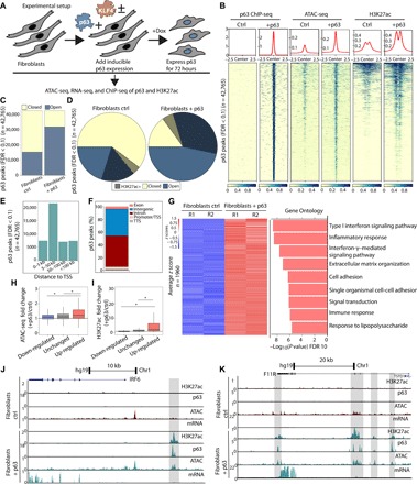
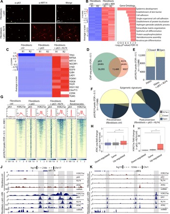
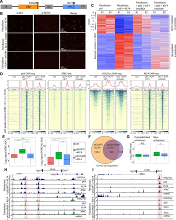
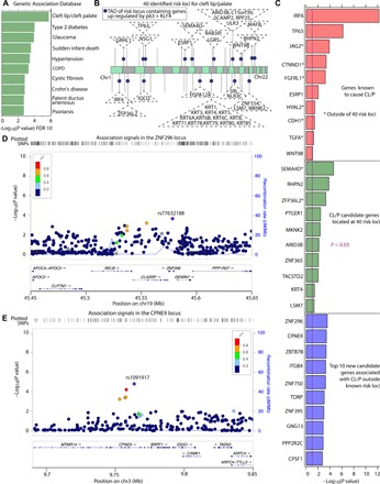
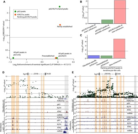
The transcription factor p63 is a key mediator of epidermal development. Point mutations in p63 in patients lead to developmental defects, including orofacial clefting. To date, knowledge on how pivotal the role of p63 is in human craniofacial development is limited. Using an inducible transdifferentiation model, combined with epigenomic sequencing and multicohort meta-analysis of genome-wide association studies data, we show that p63 establishes enhancers at craniofacial development genes to modulate their transcription. Disease-specific substitution mutation in the DNA binding domain or sterile alpha motif protein interaction domain of p63, respectively, eliminates or reduces establishment of these enhancers. We show that enhancers established by p63 are highly enriched for single-nucleotide polymorphisms associated with nonsyndromic cleft lip ± cleft palate (CL/P). These orthogonal approaches indicate a strong molecular link between p63 enhancer function and CL/P, illuminating molecular mechanisms underlying this developmental defect and revealing vital regulatory elements and new candidate causative genes.


Figures

Fig. 1
Fig. 1. p63 remodels chromatin to establish enhancers and up-regulate epithelial and inflammation genes.
(A) Experimental setup showing inducible expression of p63 and downstream epigenomic analyses. Dox, doxycycline. (B) Heatmap of p63 ChIP-seq, ATAC-seq, and H3K27ac ChIP-seq (±2.5 kb from peak center) in control fibroblasts expressing an empty vector (ctrl) and in fibroblasts expressing p63 for 72 hours. (C) Graph of called p63 peaks partitioned into open and closed chromatin according to ATAC-seq results in fibroblasts ctrl and fibroblasts + p63. (D) Pie chart depicting differences in chromatin landscape at p63 called peaks in ctrl and after ectopic expression of p63 showing clear increase in chromatin accessibility (blue, open) and H3K27ac (patterned lines). (E) Distance to nearest TSS for all p63 peaks. (F) Partitioning of p63 peaks into different genomic features. (G) Heatmap of z-scored RNA sequencing (RNA-seq) results for 1960 genes up-regulated (fold change > 1.5; FDR < 0.05) upon ectopic expression of p63 and top Gene Ontology (GO) categories showing enrichment of epithelial and inflammation categories among up-regulated genes. (H and I) Boxplots showing increased chromatin accessibility and H3K27ac at p63/KLF4 sites ±250 bp near up-regulated genes (*P < 10 × 10−10). (J) UCSC genome browser tracks showing transcriptional activation of IRF6 (interferon regulatory factor 6) and de novo H3K27ac and ATAC-seq signal at an enhancer upstream of IRF6 in fibroblasts with ectopic expression of p63 (gray highlighted box). Chr1, chromosome 1. (K) UCSC genome browser tracks showing transcriptional activation of F11R and de novo H3K27ac and ATAC-seq signal within introns of F11R in fibroblasts with ectopic expression of p63 (gray highlighted boxes).
Fig. 2
Fig. 2. p63 and KLF4 coestablish keratinocyte-specific enhancers to convert fibroblasts into keratinocyte-like cells.
(A) Immunofluorescence of p63 and KRT14 in fibroblasts + p63 and fibroblasts + p63 + KLF4 showing that KRT14 is only up-regulated when both p63 and KLF4 are expressed. (B) Heatmap of z-scored RNA-seq results for 2213 genes up-regulated (fold change > 1.5; FDR < 0.05) upon ectopic expression of p63 + KLF4 showing high enrichment of epidermal and skin-related GO categories. (C) Comparison of transcriptional regulation of the top 15 genes up-regulated in fibroblasts + p63 + KLF4 showing that both factors are required for up-regulation of keratinocyte-specific genes. (D) Venn diagram showing that p63 and KLF4 are cobound in 13,488 loci in the genome. (E) Graph of p63/KLF4 peaks partitioned into open and closed chromatin according to peaks called from ATAC-seq showing increased chromatin accessibility after 72 hours at cobound sites. Fblsts, fibroblasts. (F) Pie charts showing stark increase in chromatin accessibility and H3K27ac at p63/KLF4 peaks. (G) Heatmap showing H3K27ac enrichment flanking p63/KLF4 peaks (±2.5 kb from peak center) shared in p63 ChIP-seq of basal keratinocytes, comparing fibroblasts, fibroblasts + p63, and fibroblasts + p63 + KLF4, and reanalyzed basal keratinocyte data from (8). (H and I) Boxplots showing increased chromatin accessibility and H3K27ac at p63/KLF4 sites near up-regulated genes (*P < 10 × 10−10). (J and K) UCSC genome browser tracks showing de novo H3K27ac and ATAC-seq signal at enhancer and promoter regions close to keratinocyte genes KRT14 and IRF6, as well as transcriptional activation (gray highlighted box).
Fig. 3
Fig. 3. Disease-specific p63 mutations found in patients with craniofacial malformations show defects in enhancer establishment.
(A) Linear schematic of p63, showing domain structure and selected patient-derived mutations. TA, transactivation; OD, oligomerization domain; TID, terminal inhibitory domain. (B) Immunofluorescence of p63 and KRT14 in fibroblasts + fibroblasts + KLF4 and +WT p63, mtDBD, or mtSAM showing that KRT14 is not up-regulated when the DBD is mutated and induction of KRT14 is lower in mtSAM. (C) Heatmap of z-scored 3367 differentially expressed genes (fold change > 1.5; FDR < 0.05) between fibroblasts ctrl and fibroblasts + p63 + KLF4 showing that mtDBD has almost no change in transcriptional profile and mtSAM shows a transcriptional profile in between fibroblasts ctrl and fibroblast + WT p63 + KLF4. (D) Heatmap showing a stark reduction in chromatin accessibility (ATAC-seq signal) in mtSAM compared to WT p63, decreased H3K27ac, and retained p63 and KLF4 binding. (E) Boxplots showing no increase in chromatin accessibility for mtSAM, as well as reduced enrichment of H3K27ac flanking p63/KLF4 peaks. AUC, area under the curve; N.S., not significant. (F) Venn diagram showing that more than 1000 genes are no longer up-regulated in mtSAM + KLF4 and about 200 genes are up-regulated de novo by this mutant. (G) Boxplots showing that the RNA-seq signal of genes close to p63/KLF4 retained peaks separated into peaks at preestablished enhancers in fibroblasts and newly established enhancers after ectopic expression of p63/KLF4. (H and I) UCSC genome browser tracks showing defects (red boxes) in establishing open chromatin and inducing gene expression at FOXN1 and IRF6.
Fig. 4
Fig. 4. p63 regulates expression of critical genes associated with orofacial clefting.
(A) Genetic association database results showing that cleft lip/cleft palate is the most enriched disease category among the 2213 genes up-regulated (fold change > 1.5; FDR < 0.05) by p63 and KLF4. (B) Forty identified risk loci for cleft lip/palate indicated by black lines at the respective chromosomes with dark blue circles at topologically associated domains (TADs) containing at least one gene up-regulated by both p63 and KLF4 (respective genes in dashed triangles). (C) List showing association values of top 10 genes calculated by gene-based analysis with MAGMA as implemented in FUMA (80) in the following categories: red, genes known to cause CL/P with asterisk marking genes outside of the known 40 risk loci; green, CL/P candidate genes located at 40 risk loci with significant (P < 0.05) association to CL/P; blue, new candidate genes associated with CL/P outside of known risk loci. (D and E) Regional association plots of ZNF296 and CPNE9, showing nsCL/P-associated SNPs with nominal significant gene-based P value (<0.05) within the genes and up to 200 kb away from TSSs.
Fig. 5
Fig. 5. SNPs associated with nonsyndromic cleft lip with or without cleft palate are highly enriched in enhancers established by p63 and KLF4.
(A) GREGOR analysis showing that p63/KLF4 shared peaks (green) and newly established H3K27ac (orange) flanking these peaks are significantly (P < 0.05) enriched for SNPs associated with nsCL/P. (B) Colocalization of shared p63/KLF4 peaks at inactive (inact.)/preestablished (preestbl.)/new enhancers, with nominal significant nsCL/P-associated GWAS SNPs. The graph shows that only p63/KLF4 peaks flanked by de novo H3K27ac peaks are enriched for nsCL/P-associated SNPs. (C) Colocalization of H3K27ac peaks with nominal significant nsCL/P-associated GWAS SNPs. The graph shows that only new enhancers in converted cells flanking p63/KLF4 peaks are significantly enriched for nsCL/P-associated SNPs. (D and E) Overlay of SNPs and UCSC genome browser tracks highlighting (in orange) that p63/KLF4 peaks strongly colocalize with highly associated nsCL/P SNPs near MAFB and the known 8q24 locus; binding of both proteins at promoters is highlighted in gray.

References

    1. Yang A., Kaghad M., Wang Y., Gillett E., Fleming M. D., Dötsch V., Andrews N. C., Caput D., McKeon F., p63, a p53 homolog at 3q27–29, encodes multiple products with transactivating, death-inducing, and dominant-negative activities. Mol. Cell 2, 305–316 (1998). - PubMed
    1. Yang A., Schweitzer R., Sun D., Kaghad M., Walker N., Bronson R. T., Tabin C., Sharpe A., Caput D., Crum C., McKeon F., p63 is essential for regenerative proliferation in limb, craniofacial and epithelial development. Nature 398, 714–718 (1999). - PubMed
    1. Mills A. A., Zheng B., Wang X. J., Vogel H., Roop D. R., Bradley A., p63 is a p53 homologue required for limb and epidermal morphogenesis. Nature 398, 708–713 (1999). - PubMed
    1. Lopez-Pajares V., Yan K., Zarnegar B. J., Jameson K. L., Khavari P. A., Genetic pathways in disorders of epidermal differentiation. Trends Genet. 29, 31–40 (2013). - PMC - PubMed
    1. Celli J., Duijf P., Hamel B. C. J., Bamshad M., Kramer B., Smits A. P. T., Newbury-Ecob R., Hennekam R. C. M., van Buggenhout G., van Haeringen A., Woods C. G., van Essen A. J., de Waal R., Vriend G., Haber D. A., Yang A., McKeon F., Brunner H. G., van Bokhoven H., Heterozygous germline mutations in the p53 homolog p63 are the cause of EEC syndrome. Cell 99, 143–153 (1999). - PubMed

Publication types

MeSH terms

Supplementary concepts